Calcium »
PDB 4qn5-4r90 »
4r12 »
Calcium in PDB 4r12: Crystal Structure of the Gamma-Secretase Component Nicastrin
Protein crystallography data
The structure of Crystal Structure of the Gamma-Secretase Component Nicastrin, PDB code: 4r12
was solved by
T.Xie,
C.Yan,
R.Zhou,
Y.Zhao,
L.Sun,
G.Yang,
P.Lu,
D.Ma,
Y.Shi,
with X-Ray Crystallography technique. A brief refinement statistics is given in the table below:
| Resolution Low / High (Å) | 36.08 / 1.95 |
| Space group | P 41 21 2 |
| Cell size a, b, c (Å), α, β, γ (°) | 65.584, 65.584, 344.475, 90.00, 90.00, 90.00 |
| R / Rfree (%) | 17.9 / 20.5 |
Calcium Binding Sites:
The binding sites of Calcium atom in the Crystal Structure of the Gamma-Secretase Component Nicastrin
(pdb code 4r12). This binding sites where shown within
5.0 Angstroms radius around Calcium atom.
In total only one binding site of Calcium was determined in the Crystal Structure of the Gamma-Secretase Component Nicastrin, PDB code: 4r12:
In total only one binding site of Calcium was determined in the Crystal Structure of the Gamma-Secretase Component Nicastrin, PDB code: 4r12:
Calcium binding site 1 out of 1 in 4r12
Go back to
Calcium binding site 1 out
of 1 in the Crystal Structure of the Gamma-Secretase Component Nicastrin
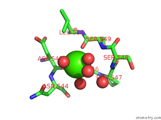
Mono view
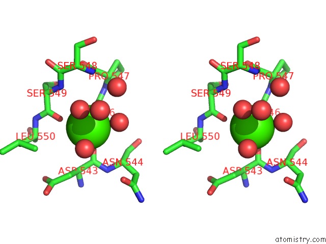
Stereo pair view
Mono view
Stereo pair view
A full contact list of Calcium with other atoms in the Ca binding
site number 1 of Crystal Structure of the Gamma-Secretase Component Nicastrin within 5.0Å range:
|
Reference:
T.Xie,
C.Yan,
R.Zhou,
Y.Zhao,
L.Sun,
G.Yang,
P.Lu,
D.Ma,
Y.Shi.
Crystal Structure of the Gamma-Secretase Component Nicastrin Proc.Natl.Acad.Sci.Usa 2014.
ISSN: ESSN 1091-6490
PubMed: 25197054
DOI: 10.1073/PNAS.1414837111
Page generated: Sun Jul 14 12:33:56 2024
ISSN: ESSN 1091-6490
PubMed: 25197054
DOI: 10.1073/PNAS.1414837111
Last articles
Zn in 9J0NZn in 9J0O
Zn in 9J0P
Zn in 9FJX
Zn in 9EKB
Zn in 9C0F
Zn in 9CAH
Zn in 9CH0
Zn in 9CH3
Zn in 9CH1