Calcium »
PDB 4qn5-4r90 »
4r49 »
Calcium in PDB 4r49: Racemic Crystal Structure of A Calcium-Bound B-Dna Duplex
Protein crystallography data
The structure of Racemic Crystal Structure of A Calcium-Bound B-Dna Duplex, PDB code: 4r49
was solved by
P.K.Mandal,
G.W.Collie,
B.Kauffmann,
I.Huc,
with X-Ray Crystallography technique. A brief refinement statistics is given in the table below:
| Resolution Low / High (Å) | 34.25 / 1.28 |
| Space group | P -1 |
| Cell size a, b, c (Å), α, β, γ (°) | 23.263, 33.604, 36.820, 103.32, 101.02, 109.78 |
| R / Rfree (%) | 21.7 / 26 |
Other elements in 4r49:
The structure of Racemic Crystal Structure of A Calcium-Bound B-Dna Duplex also contains other interesting chemical elements:
| Sodium | (Na) | 2 atoms |
Calcium Binding Sites:
The binding sites of Calcium atom in the Racemic Crystal Structure of A Calcium-Bound B-Dna Duplex
(pdb code 4r49). This binding sites where shown within
5.0 Angstroms radius around Calcium atom.
In total 7 binding sites of Calcium where determined in the Racemic Crystal Structure of A Calcium-Bound B-Dna Duplex, PDB code: 4r49:
Jump to Calcium binding site number: 1; 2; 3; 4; 5; 6; 7;
In total 7 binding sites of Calcium where determined in the Racemic Crystal Structure of A Calcium-Bound B-Dna Duplex, PDB code: 4r49:
Jump to Calcium binding site number: 1; 2; 3; 4; 5; 6; 7;
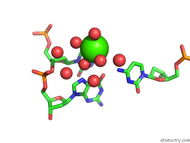
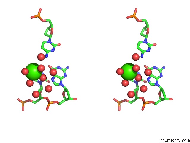
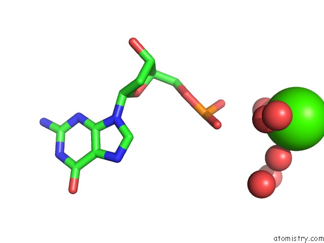
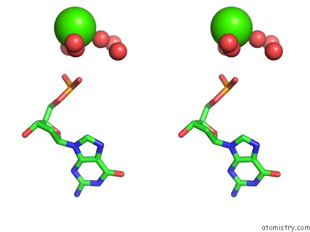
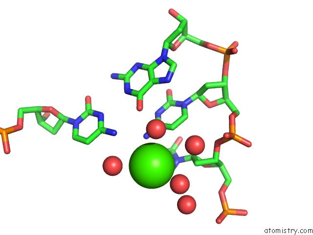
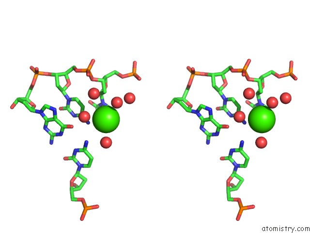
Calcium binding site 1 out of 7 in 4r49
Go back to
Calcium binding site 1 out
of 7 in the Racemic Crystal Structure of A Calcium-Bound B-Dna Duplex
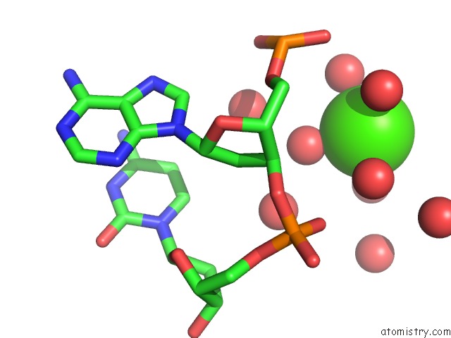
Mono view
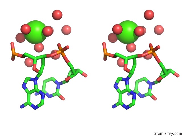
Stereo pair view
Mono view
Stereo pair view
A full contact list of Calcium with other atoms in the Ca binding
site number 1 of Racemic Crystal Structure of A Calcium-Bound B-Dna Duplex within 5.0Å range:
|
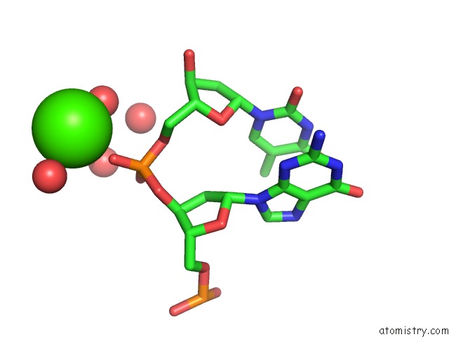
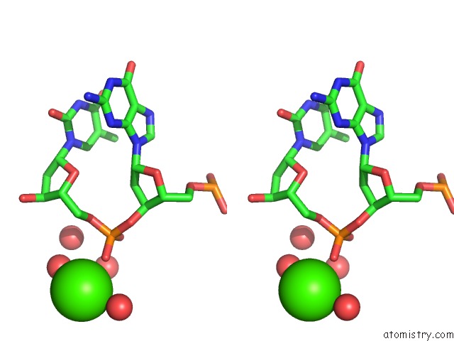
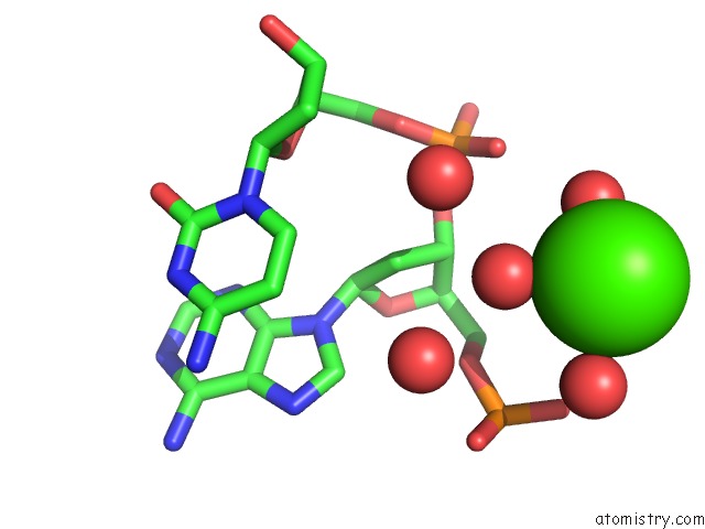
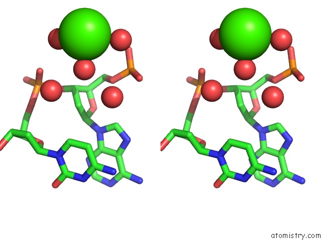
Calcium binding site 2 out of 7 in 4r49
Go back to
Calcium binding site 2 out
of 7 in the Racemic Crystal Structure of A Calcium-Bound B-Dna Duplex
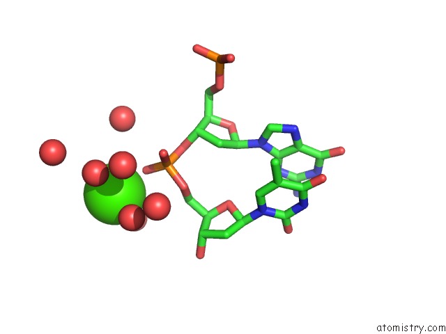
Mono view
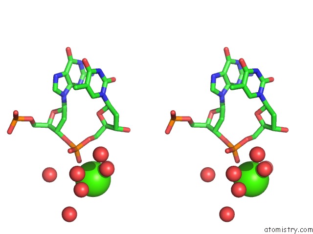
Stereo pair view
Mono view
Stereo pair view
A full contact list of Calcium with other atoms in the Ca binding
site number 2 of Racemic Crystal Structure of A Calcium-Bound B-Dna Duplex within 5.0Å range:
|
Calcium binding site 3 out of 7 in 4r49
Go back to
Calcium binding site 3 out
of 7 in the Racemic Crystal Structure of A Calcium-Bound B-Dna Duplex
Mono view
Stereo pair view
Mono view
Stereo pair view
A full contact list of Calcium with other atoms in the Ca binding
site number 3 of Racemic Crystal Structure of A Calcium-Bound B-Dna Duplex within 5.0Å range:
|
Calcium binding site 4 out of 7 in 4r49
Go back to
Calcium binding site 4 out
of 7 in the Racemic Crystal Structure of A Calcium-Bound B-Dna Duplex
Mono view
Stereo pair view
Mono view
Stereo pair view
A full contact list of Calcium with other atoms in the Ca binding
site number 4 of Racemic Crystal Structure of A Calcium-Bound B-Dna Duplex within 5.0Å range:
|
Calcium binding site 5 out of 7 in 4r49
Go back to
Calcium binding site 5 out
of 7 in the Racemic Crystal Structure of A Calcium-Bound B-Dna Duplex
Mono view
Stereo pair view
Mono view
Stereo pair view
A full contact list of Calcium with other atoms in the Ca binding
site number 5 of Racemic Crystal Structure of A Calcium-Bound B-Dna Duplex within 5.0Å range:
|
Calcium binding site 6 out of 7 in 4r49
Go back to
Calcium binding site 6 out
of 7 in the Racemic Crystal Structure of A Calcium-Bound B-Dna Duplex
Mono view
Stereo pair view
Mono view
Stereo pair view
A full contact list of Calcium with other atoms in the Ca binding
site number 6 of Racemic Crystal Structure of A Calcium-Bound B-Dna Duplex within 5.0Å range:
|
Calcium binding site 7 out of 7 in 4r49
Go back to
Calcium binding site 7 out
of 7 in the Racemic Crystal Structure of A Calcium-Bound B-Dna Duplex
Mono view
Stereo pair view
Mono view
Stereo pair view
A full contact list of Calcium with other atoms in the Ca binding
site number 7 of Racemic Crystal Structure of A Calcium-Bound B-Dna Duplex within 5.0Å range:
|
Reference:
P.K.Mandal,
G.W.Collie,
B.Kauffmann,
I.Huc.
Racemic Dna Crystallography. Angew.Chem.Int.Ed.Engl. 2014.
ISSN: ESSN 1521-3773
PubMed: 25358289
DOI: 10.1002/ANIE.201409014
Page generated: Sun Jul 14 12:36:05 2024
ISSN: ESSN 1521-3773
PubMed: 25358289
DOI: 10.1002/ANIE.201409014
Last articles
Zn in 9J0NZn in 9J0O
Zn in 9J0P
Zn in 9FJX
Zn in 9EKB
Zn in 9C0F
Zn in 9CAH
Zn in 9CH0
Zn in 9CH3
Zn in 9CH1