Calcium »
PDB 4r9j-4rvy »
4rfu »
Calcium in PDB 4rfu: Crystal Structure of Truncated P-Domain From Grouper Nervous Necrosis Virus Capsid Protein at 1.2A
Protein crystallography data
The structure of Crystal Structure of Truncated P-Domain From Grouper Nervous Necrosis Virus Capsid Protein at 1.2A, PDB code: 4rfu
was solved by
N.C.Chen,
C.J.Chen,
M.Yoshimura,
H.H.Guan,
T.Y.Chen,
with X-Ray Crystallography technique. A brief refinement statistics is given in the table below:
| Resolution Low / High (Å) | 20.76 / 1.20 |
| Space group | P 21 21 21 |
| Cell size a, b, c (Å), α, β, γ (°) | 64.982, 83.458, 85.577, 90.00, 90.00, 90.00 |
| R / Rfree (%) | 17.5 / 18.1 |
Calcium Binding Sites:
The binding sites of Calcium atom in the Crystal Structure of Truncated P-Domain From Grouper Nervous Necrosis Virus Capsid Protein at 1.2A
(pdb code 4rfu). This binding sites where shown within
5.0 Angstroms radius around Calcium atom.
In total 2 binding sites of Calcium where determined in the Crystal Structure of Truncated P-Domain From Grouper Nervous Necrosis Virus Capsid Protein at 1.2A, PDB code: 4rfu:
Jump to Calcium binding site number: 1; 2;
In total 2 binding sites of Calcium where determined in the Crystal Structure of Truncated P-Domain From Grouper Nervous Necrosis Virus Capsid Protein at 1.2A, PDB code: 4rfu:
Jump to Calcium binding site number: 1; 2;
Calcium binding site 1 out of 2 in 4rfu
Go back to
Calcium binding site 1 out
of 2 in the Crystal Structure of Truncated P-Domain From Grouper Nervous Necrosis Virus Capsid Protein at 1.2A
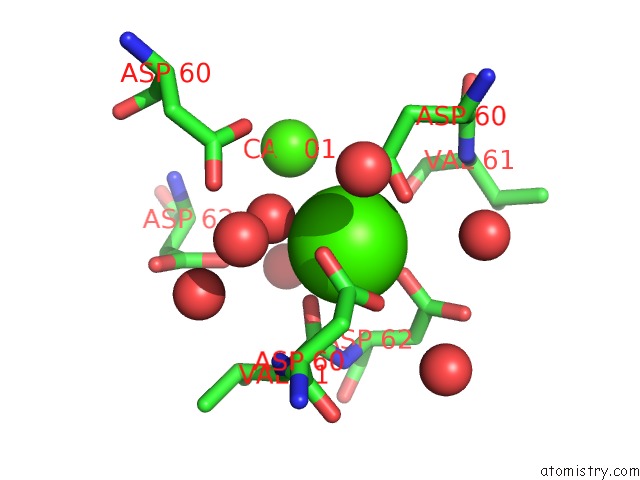
Mono view
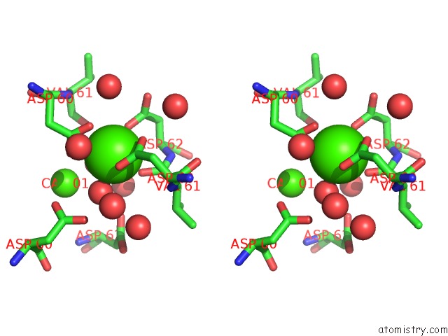
Stereo pair view
Mono view
Stereo pair view
A full contact list of Calcium with other atoms in the Ca binding
site number 1 of Crystal Structure of Truncated P-Domain From Grouper Nervous Necrosis Virus Capsid Protein at 1.2A within 5.0Å range:
|
Calcium binding site 2 out of 2 in 4rfu
Go back to
Calcium binding site 2 out
of 2 in the Crystal Structure of Truncated P-Domain From Grouper Nervous Necrosis Virus Capsid Protein at 1.2A
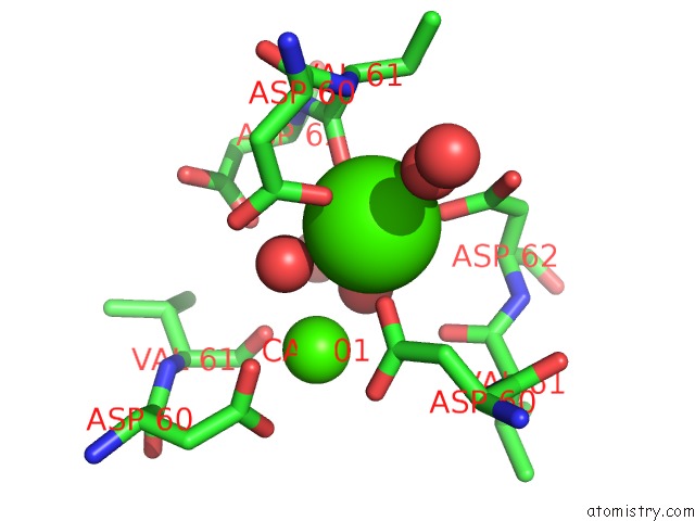
Mono view
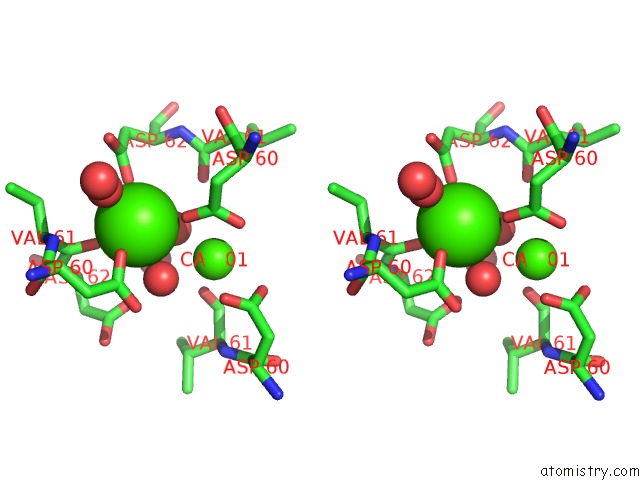
Stereo pair view
Mono view
Stereo pair view
A full contact list of Calcium with other atoms in the Ca binding
site number 2 of Crystal Structure of Truncated P-Domain From Grouper Nervous Necrosis Virus Capsid Protein at 1.2A within 5.0Å range:
|
Reference:
N.C.Chen,
M.Yoshimura,
H.H.Guan,
T.Y.Wang,
Y.Misumi,
C.C.Lin,
P.Chuankhayan,
A.Nakagawa,
S.I.Chan,
T.Tsukihara,
T.Y.Chen,
C.J.Chen.
Crystal Structures of A Piscine Betanodavirus: Mechanisms of Capsid Assembly and Viral Infection Plos Pathog. V. 11 05203 2015.
ISSN: ISSN 1553-7366
PubMed: 26491970
DOI: 10.1371/JOURNAL.PPAT.1005203
Page generated: Sun Jul 14 12:40:15 2024
ISSN: ISSN 1553-7366
PubMed: 26491970
DOI: 10.1371/JOURNAL.PPAT.1005203
Last articles
Zn in 9JYWZn in 9IR4
Zn in 9IR3
Zn in 9GMX
Zn in 9GMW
Zn in 9JEJ
Zn in 9ERF
Zn in 9ERE
Zn in 9EGV
Zn in 9EGW