Calcium »
PDB 4ry0-4twe »
4tv5 »
Calcium in PDB 4tv5: Crystal Structure of Citrate Synthase Sbng
Protein crystallography data
The structure of Crystal Structure of Citrate Synthase Sbng, PDB code: 4tv5
was solved by
M.J.Kobylarz,
J.C.Grigg,
M.E.P.Murphy,
with X-Ray Crystallography technique. A brief refinement statistics is given in the table below:
| Resolution Low / High (Å) | 76.72 / 1.85 |
| Space group | P 3 1 2 |
| Cell size a, b, c (Å), α, β, γ (°) | 74.290, 74.290, 76.720, 90.00, 90.00, 120.00 |
| R / Rfree (%) | 19.7 / 21.1 |
Calcium Binding Sites:
The binding sites of Calcium atom in the Crystal Structure of Citrate Synthase Sbng
(pdb code 4tv5). This binding sites where shown within
5.0 Angstroms radius around Calcium atom.
In total 2 binding sites of Calcium where determined in the Crystal Structure of Citrate Synthase Sbng, PDB code: 4tv5:
Jump to Calcium binding site number: 1; 2;
In total 2 binding sites of Calcium where determined in the Crystal Structure of Citrate Synthase Sbng, PDB code: 4tv5:
Jump to Calcium binding site number: 1; 2;
Calcium binding site 1 out of 2 in 4tv5
Go back to
Calcium binding site 1 out
of 2 in the Crystal Structure of Citrate Synthase Sbng
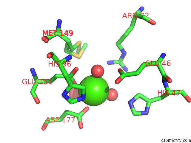
Mono view
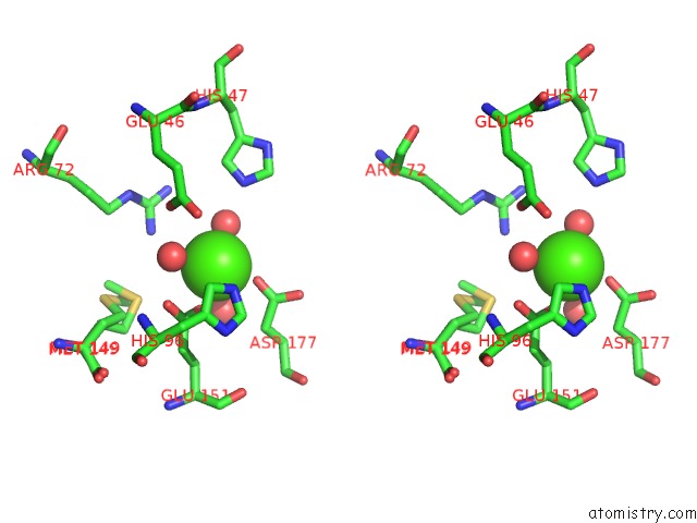
Stereo pair view
Mono view
Stereo pair view
A full contact list of Calcium with other atoms in the Ca binding
site number 1 of Crystal Structure of Citrate Synthase Sbng within 5.0Å range:
|
Calcium binding site 2 out of 2 in 4tv5
Go back to
Calcium binding site 2 out
of 2 in the Crystal Structure of Citrate Synthase Sbng
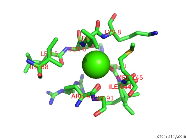
Mono view
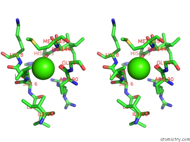
Stereo pair view
Mono view
Stereo pair view
A full contact list of Calcium with other atoms in the Ca binding
site number 2 of Crystal Structure of Citrate Synthase Sbng within 5.0Å range:
|
Reference:
M.J.Kobylarz,
J.C.Grigg,
J.R.Sheldon,
D.E.Heinrichs,
M.E.Murphy.
Sbng, A Citrate Synthase in Staphylococcus Aureus: A New Fold on An Old Enzyme. J.Biol.Chem. 2014.
ISSN: ESSN 1083-351X
PubMed: 25336653
DOI: 10.1074/JBC.M114.603175
Page generated: Sun Jul 14 13:31:12 2024
ISSN: ESSN 1083-351X
PubMed: 25336653
DOI: 10.1074/JBC.M114.603175
Last articles
Zn in 9J0NZn in 9J0O
Zn in 9J0P
Zn in 9FJX
Zn in 9EKB
Zn in 9C0F
Zn in 9CAH
Zn in 9CH0
Zn in 9CH3
Zn in 9CH1