Calcium »
PDB 4ry0-4twe »
4twe »
Calcium in PDB 4twe: Structure of Ligand-Free N-Acetylated-Alpha-Linked-Acidic-Dipeptidase Like Protein (Naaladasel)
Enzymatic activity of Structure of Ligand-Free N-Acetylated-Alpha-Linked-Acidic-Dipeptidase Like Protein (Naaladasel)
All present enzymatic activity of Structure of Ligand-Free N-Acetylated-Alpha-Linked-Acidic-Dipeptidase Like Protein (Naaladasel):
3.4.17.21;
3.4.17.21;
Protein crystallography data
The structure of Structure of Ligand-Free N-Acetylated-Alpha-Linked-Acidic-Dipeptidase Like Protein (Naaladasel), PDB code: 4twe
was solved by
J.Tykvart,
C.Barinka,
J.Lubkowski,
P.Sacha,
J.Konvalinka,
with X-Ray Crystallography technique. A brief refinement statistics is given in the table below:
| Resolution Low / High (Å) | 29.14 / 1.75 |
| Space group | I 2 2 2 |
| Cell size a, b, c (Å), α, β, γ (°) | 98.512, 174.844, 208.034, 90.00, 90.00, 90.00 |
| R / Rfree (%) | 16.7 / 18.8 |
Other elements in 4twe:
The structure of Structure of Ligand-Free N-Acetylated-Alpha-Linked-Acidic-Dipeptidase Like Protein (Naaladasel) also contains other interesting chemical elements:
| Zinc | (Zn) | 4 atoms |
Calcium Binding Sites:
The binding sites of Calcium atom in the Structure of Ligand-Free N-Acetylated-Alpha-Linked-Acidic-Dipeptidase Like Protein (Naaladasel)
(pdb code 4twe). This binding sites where shown within
5.0 Angstroms radius around Calcium atom.
In total 2 binding sites of Calcium where determined in the Structure of Ligand-Free N-Acetylated-Alpha-Linked-Acidic-Dipeptidase Like Protein (Naaladasel), PDB code: 4twe:
Jump to Calcium binding site number: 1; 2;
In total 2 binding sites of Calcium where determined in the Structure of Ligand-Free N-Acetylated-Alpha-Linked-Acidic-Dipeptidase Like Protein (Naaladasel), PDB code: 4twe:
Jump to Calcium binding site number: 1; 2;
Calcium binding site 1 out of 2 in 4twe
Go back to
Calcium binding site 1 out
of 2 in the Structure of Ligand-Free N-Acetylated-Alpha-Linked-Acidic-Dipeptidase Like Protein (Naaladasel)
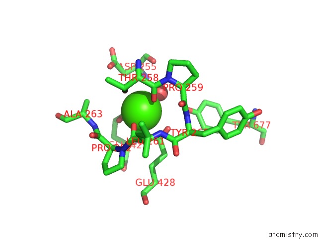
Mono view
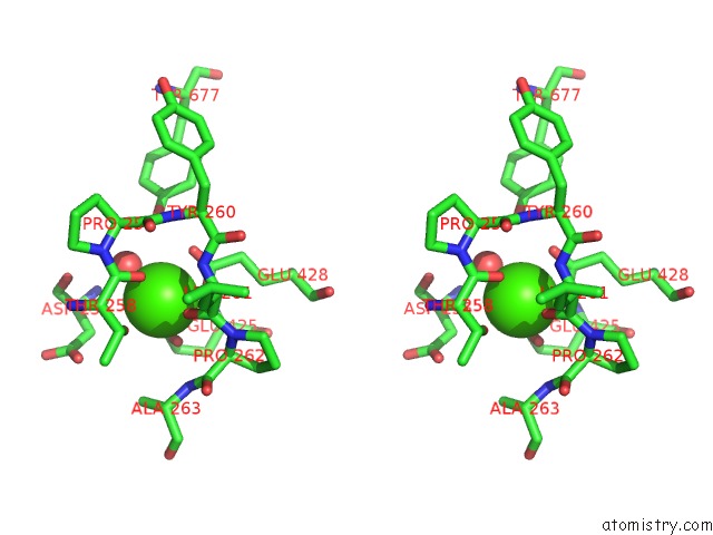
Stereo pair view
Mono view
Stereo pair view
A full contact list of Calcium with other atoms in the Ca binding
site number 1 of Structure of Ligand-Free N-Acetylated-Alpha-Linked-Acidic-Dipeptidase Like Protein (Naaladasel) within 5.0Å range:
|
Calcium binding site 2 out of 2 in 4twe
Go back to
Calcium binding site 2 out
of 2 in the Structure of Ligand-Free N-Acetylated-Alpha-Linked-Acidic-Dipeptidase Like Protein (Naaladasel)
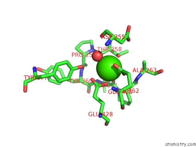
Mono view
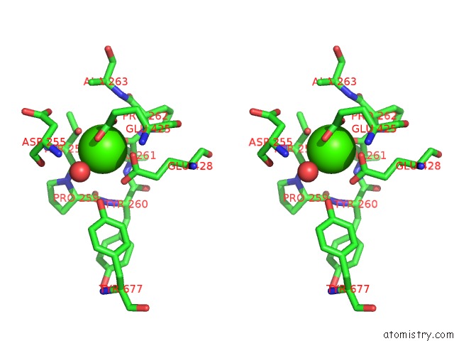
Stereo pair view
Mono view
Stereo pair view
A full contact list of Calcium with other atoms in the Ca binding
site number 2 of Structure of Ligand-Free N-Acetylated-Alpha-Linked-Acidic-Dipeptidase Like Protein (Naaladasel) within 5.0Å range:
|
Reference:
J.Tykvart,
C.Barinka,
M.Svoboda,
V.Navratil,
R.Soucek,
M.Hubalek,
M.Hradilek,
P.Sacha,
J.Lubkowski,
J.Konvalinka.
Structural and Biochemical Characterization of A Novel Aminopeptidase From Human Intestine J.Biol.Chem. 2015.
ISSN: ESSN 1083-351X
DOI: 10.1074/JBC.M114.628149
Page generated: Sun Jul 14 13:33:06 2024
ISSN: ESSN 1083-351X
DOI: 10.1074/JBC.M114.628149
Last articles
Zn in 9MJ5Zn in 9HNW
Zn in 9G0L
Zn in 9FNE
Zn in 9DZN
Zn in 9E0I
Zn in 9D32
Zn in 9DAK
Zn in 8ZXC
Zn in 8ZUF