Calcium »
PDB 4tx8-4uix »
4u65 »
Calcium in PDB 4u65: Structure of the Periplasmic Output Domain of the Legionella Pneumophila Lapd Ortholog CDGS9 in Complex with Pseudomonas Fluorescens Lapg
Protein crystallography data
The structure of Structure of the Periplasmic Output Domain of the Legionella Pneumophila Lapd Ortholog CDGS9 in Complex with Pseudomonas Fluorescens Lapg, PDB code: 4u65
was solved by
D.Chatterjee,
R.B.Cooley,
C.D.Boyd,
R.A.Mehl,
G.A.O'toole,
H.S.Sondermann,
with X-Ray Crystallography technique. A brief refinement statistics is given in the table below:
| Resolution Low / High (Å) | 44.06 / 2.10 |
| Space group | P 1 21 1 |
| Cell size a, b, c (Å), α, β, γ (°) | 75.854, 73.674, 88.223, 90.00, 92.88, 90.00 |
| R / Rfree (%) | 17 / 21.1 |
Calcium Binding Sites:
The binding sites of Calcium atom in the Structure of the Periplasmic Output Domain of the Legionella Pneumophila Lapd Ortholog CDGS9 in Complex with Pseudomonas Fluorescens Lapg
(pdb code 4u65). This binding sites where shown within
5.0 Angstroms radius around Calcium atom.
In total 4 binding sites of Calcium where determined in the Structure of the Periplasmic Output Domain of the Legionella Pneumophila Lapd Ortholog CDGS9 in Complex with Pseudomonas Fluorescens Lapg, PDB code: 4u65:
Jump to Calcium binding site number: 1; 2; 3; 4;
In total 4 binding sites of Calcium where determined in the Structure of the Periplasmic Output Domain of the Legionella Pneumophila Lapd Ortholog CDGS9 in Complex with Pseudomonas Fluorescens Lapg, PDB code: 4u65:
Jump to Calcium binding site number: 1; 2; 3; 4;
Calcium binding site 1 out of 4 in 4u65
Go back to
Calcium binding site 1 out
of 4 in the Structure of the Periplasmic Output Domain of the Legionella Pneumophila Lapd Ortholog CDGS9 in Complex with Pseudomonas Fluorescens Lapg
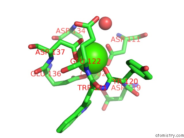
Mono view

Stereo pair view
Mono view
Stereo pair view
A full contact list of Calcium with other atoms in the Ca binding
site number 1 of Structure of the Periplasmic Output Domain of the Legionella Pneumophila Lapd Ortholog CDGS9 in Complex with Pseudomonas Fluorescens Lapg within 5.0Å range:
|
Calcium binding site 2 out of 4 in 4u65
Go back to
Calcium binding site 2 out
of 4 in the Structure of the Periplasmic Output Domain of the Legionella Pneumophila Lapd Ortholog CDGS9 in Complex with Pseudomonas Fluorescens Lapg
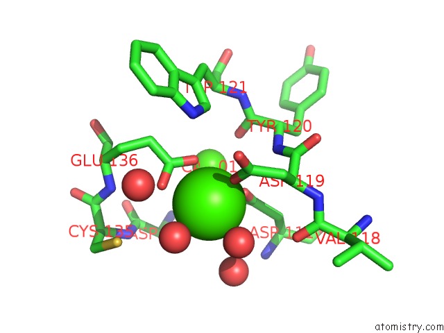
Mono view
Stereo pair view
Mono view
Stereo pair view
A full contact list of Calcium with other atoms in the Ca binding
site number 2 of Structure of the Periplasmic Output Domain of the Legionella Pneumophila Lapd Ortholog CDGS9 in Complex with Pseudomonas Fluorescens Lapg within 5.0Å range:
|
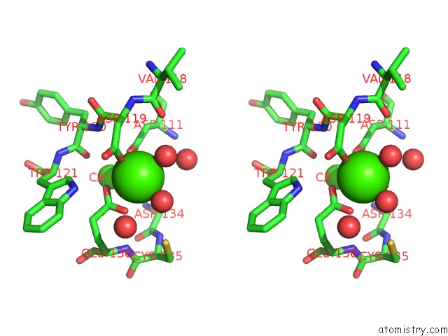
Calcium binding site 3 out of 4 in 4u65
Go back to
Calcium binding site 3 out
of 4 in the Structure of the Periplasmic Output Domain of the Legionella Pneumophila Lapd Ortholog CDGS9 in Complex with Pseudomonas Fluorescens Lapg

Mono view

Stereo pair view
Mono view
Stereo pair view
A full contact list of Calcium with other atoms in the Ca binding
site number 3 of Structure of the Periplasmic Output Domain of the Legionella Pneumophila Lapd Ortholog CDGS9 in Complex with Pseudomonas Fluorescens Lapg within 5.0Å range:
|
Calcium binding site 4 out of 4 in 4u65
Go back to
Calcium binding site 4 out
of 4 in the Structure of the Periplasmic Output Domain of the Legionella Pneumophila Lapd Ortholog CDGS9 in Complex with Pseudomonas Fluorescens Lapg
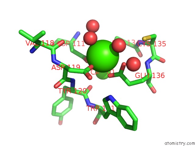
Mono view

Stereo pair view
Mono view
Stereo pair view
A full contact list of Calcium with other atoms in the Ca binding
site number 4 of Structure of the Periplasmic Output Domain of the Legionella Pneumophila Lapd Ortholog CDGS9 in Complex with Pseudomonas Fluorescens Lapg within 5.0Å range:
|
Reference:
D.Chatterjee,
R.B.Cooley,
C.D.Boyd,
R.A.Mehl,
G.A.O'toole,
H.Sondermann.
Mechanistic Insight Into the Conserved Allosteric Regulation of Periplasmic Proteolysis By the Signaling Molecule Cyclic-Di-Gmp. Elife V. 3 03650 2014.
ISSN: ESSN 2050-084X
PubMed: 25182848
DOI: 10.7554/ELIFE.03650
Page generated: Sun Jul 14 13:36:01 2024
ISSN: ESSN 2050-084X
PubMed: 25182848
DOI: 10.7554/ELIFE.03650
Last articles
Zn in 9J0NZn in 9J0O
Zn in 9J0P
Zn in 9FJX
Zn in 9EKB
Zn in 9C0F
Zn in 9CAH
Zn in 9CH0
Zn in 9CH3
Zn in 9CH1