Calcium »
PDB 4tx8-4uix »
4uap »
Calcium in PDB 4uap: X-Ray Structure of GH31 CBM32-2 Bound to Galnac
Protein crystallography data
The structure of X-Ray Structure of GH31 CBM32-2 Bound to Galnac, PDB code: 4uap
was solved by
J.M.Grondin,
K.Abe,
A.B.Boraston,
S.P.Smith,
with X-Ray Crystallography technique. A brief refinement statistics is given in the table below:
| Resolution Low / High (Å) | 38.41 / 2.00 |
| Space group | P 2 21 21 |
| Cell size a, b, c (Å), α, β, γ (°) | 48.300, 84.750, 86.170, 90.00, 90.00, 90.00 |
| R / Rfree (%) | 14.5 / 19.2 |
Calcium Binding Sites:
The binding sites of Calcium atom in the X-Ray Structure of GH31 CBM32-2 Bound to Galnac
(pdb code 4uap). This binding sites where shown within
5.0 Angstroms radius around Calcium atom.
In total 2 binding sites of Calcium where determined in the X-Ray Structure of GH31 CBM32-2 Bound to Galnac, PDB code: 4uap:
Jump to Calcium binding site number: 1; 2;
In total 2 binding sites of Calcium where determined in the X-Ray Structure of GH31 CBM32-2 Bound to Galnac, PDB code: 4uap:
Jump to Calcium binding site number: 1; 2;
Calcium binding site 1 out of 2 in 4uap
Go back to
Calcium binding site 1 out
of 2 in the X-Ray Structure of GH31 CBM32-2 Bound to Galnac
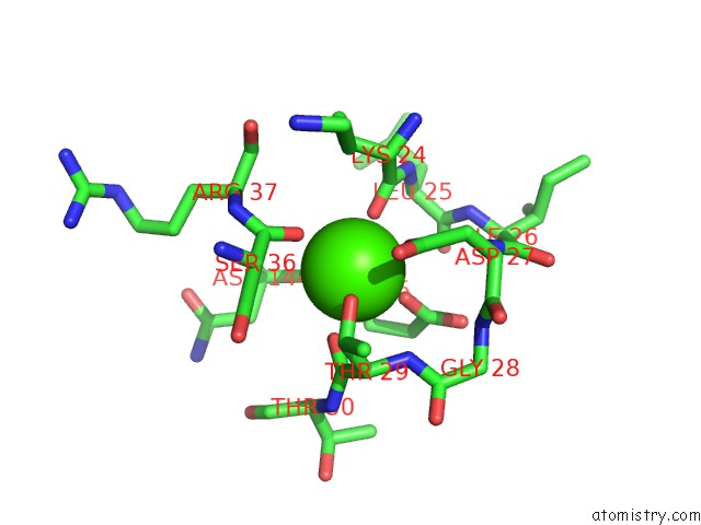
Mono view
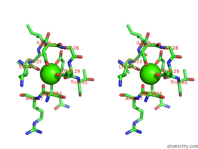
Stereo pair view
Mono view
Stereo pair view
A full contact list of Calcium with other atoms in the Ca binding
site number 1 of X-Ray Structure of GH31 CBM32-2 Bound to Galnac within 5.0Å range:
|
Calcium binding site 2 out of 2 in 4uap
Go back to
Calcium binding site 2 out
of 2 in the X-Ray Structure of GH31 CBM32-2 Bound to Galnac
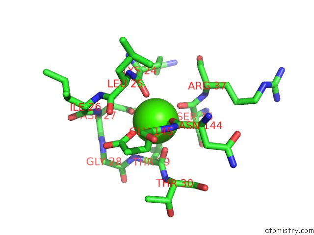
Mono view
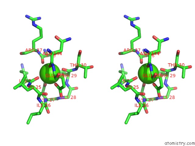
Stereo pair view
Mono view
Stereo pair view
A full contact list of Calcium with other atoms in the Ca binding
site number 2 of X-Ray Structure of GH31 CBM32-2 Bound to Galnac within 5.0Å range:
|
Reference:
J.M.Grondin,
D.Duan,
A.C.Kirlin,
K.T.Abe,
S.Chitayat,
H.L.Spencer,
C.Spencer,
A.Campigotto,
S.Houliston,
C.H.Arrowsmith,
J.S.Allingham,
A.B.Boraston,
S.P.Smith.
Diverse Modes of Galacto-Specific Carbohydrate Recognition By A Family 31 Glycoside Hydrolase From Clostridium Perfringens. Plos One V. 12 71606 2017.
ISSN: ESSN 1932-6203
PubMed: 28158290
DOI: 10.1371/JOURNAL.PONE.0171606
Page generated: Sun Jul 14 13:38:31 2024
ISSN: ESSN 1932-6203
PubMed: 28158290
DOI: 10.1371/JOURNAL.PONE.0171606
Last articles
Zn in 9J0NZn in 9J0O
Zn in 9J0P
Zn in 9FJX
Zn in 9EKB
Zn in 9C0F
Zn in 9CAH
Zn in 9CH0
Zn in 9CH3
Zn in 9CH1