Calcium »
PDB 4tx8-4uix »
4uaw »
Calcium in PDB 4uaw: Dna Polymerase Beta Substrate Complex with A Templating Adenine and Incoming 8-Oxodgtp, 0 S
Enzymatic activity of Dna Polymerase Beta Substrate Complex with A Templating Adenine and Incoming 8-Oxodgtp, 0 S
All present enzymatic activity of Dna Polymerase Beta Substrate Complex with A Templating Adenine and Incoming 8-Oxodgtp, 0 S:
2.7.7.7;
2.7.7.7;
Protein crystallography data
The structure of Dna Polymerase Beta Substrate Complex with A Templating Adenine and Incoming 8-Oxodgtp, 0 S, PDB code: 4uaw
was solved by
B.D.Freudenthal,
S.H.Wilson,
W.A.Beard,
with X-Ray Crystallography technique. A brief refinement statistics is given in the table below:
| Resolution Low / High (Å) | 37.73 / 1.90 |
| Space group | P 1 21 1 |
| Cell size a, b, c (Å), α, β, γ (°) | 50.932, 79.929, 55.515, 90.00, 107.59, 90.00 |
| R / Rfree (%) | 18.3 / 24 |
Other elements in 4uaw:
The structure of Dna Polymerase Beta Substrate Complex with A Templating Adenine and Incoming 8-Oxodgtp, 0 S also contains other interesting chemical elements:
| Sodium | (Na) | 2 atoms |
Calcium Binding Sites:
The binding sites of Calcium atom in the Dna Polymerase Beta Substrate Complex with A Templating Adenine and Incoming 8-Oxodgtp, 0 S
(pdb code 4uaw). This binding sites where shown within
5.0 Angstroms radius around Calcium atom.
In total 2 binding sites of Calcium where determined in the Dna Polymerase Beta Substrate Complex with A Templating Adenine and Incoming 8-Oxodgtp, 0 S, PDB code: 4uaw:
Jump to Calcium binding site number: 1; 2;
In total 2 binding sites of Calcium where determined in the Dna Polymerase Beta Substrate Complex with A Templating Adenine and Incoming 8-Oxodgtp, 0 S, PDB code: 4uaw:
Jump to Calcium binding site number: 1; 2;
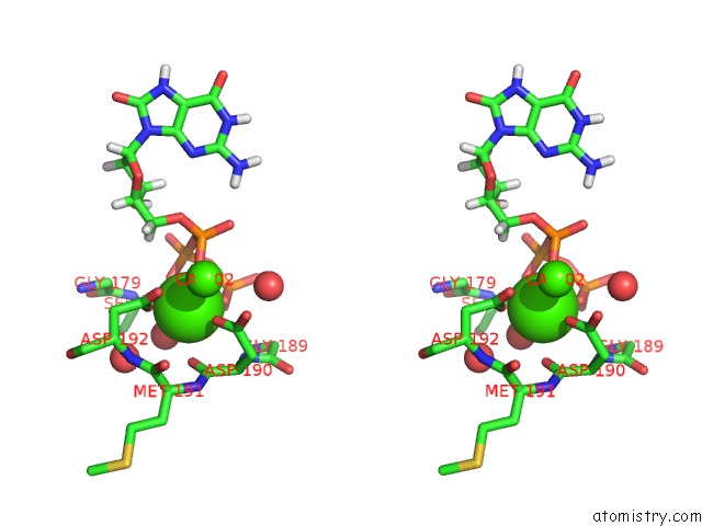
Calcium binding site 1 out of 2 in 4uaw
Go back to
Calcium binding site 1 out
of 2 in the Dna Polymerase Beta Substrate Complex with A Templating Adenine and Incoming 8-Oxodgtp, 0 S

Mono view

Stereo pair view
Mono view
Stereo pair view
A full contact list of Calcium with other atoms in the Ca binding
site number 1 of Dna Polymerase Beta Substrate Complex with A Templating Adenine and Incoming 8-Oxodgtp, 0 S within 5.0Å range:
|
Calcium binding site 2 out of 2 in 4uaw
Go back to
Calcium binding site 2 out
of 2 in the Dna Polymerase Beta Substrate Complex with A Templating Adenine and Incoming 8-Oxodgtp, 0 S

Mono view
Stereo pair view
Mono view
Stereo pair view
A full contact list of Calcium with other atoms in the Ca binding
site number 2 of Dna Polymerase Beta Substrate Complex with A Templating Adenine and Incoming 8-Oxodgtp, 0 S within 5.0Å range:
|
Reference:
B.D.Freudenthal,
W.A.Beard,
L.Perera,
D.D.Shock,
T.Kim,
T.Schlick,
S.H.Wilson.
Uncovering the Polymerase-Induced Cytotoxicty of An Oxidized Nucleotide Nature 2014.
ISSN: ESSN 1476-4687
DOI: 10.1038/NATURE13886
Page generated: Sun Jul 14 13:38:58 2024
ISSN: ESSN 1476-4687
DOI: 10.1038/NATURE13886
Last articles
Zn in 9J0NZn in 9J0O
Zn in 9J0P
Zn in 9FJX
Zn in 9EKB
Zn in 9C0F
Zn in 9CAH
Zn in 9CH0
Zn in 9CH3
Zn in 9CH1