Calcium »
PDB 4tx8-4uix »
4ufc »
Calcium in PDB 4ufc: Crystal Structure of the GH95 Enzyme BACOVA_03438
Protein crystallography data
The structure of Crystal Structure of the GH95 Enzyme BACOVA_03438, PDB code: 4ufc
was solved by
A.Rogowski,
J.A.Briggs,
J.C.Mortimer,
T.Tryfona,
N.Terrapon,
E.C.Lowe,
A.Basle,
C.Morland,
A.M.Day,
H.Zheng,
T.E.Rogers,
P.Thompson,
A.R.Hawkins,
M.P.Yadav,
B.Henrissat,
E.C.Martens,
P.Dupree,
H.J.Gilbert,
D.N.Bolam,
with X-Ray Crystallography technique. A brief refinement statistics is given in the table below:
| Resolution Low / High (Å) | 134.93 / 2.81 |
| Space group | P 21 21 21 |
| Cell size a, b, c (Å), α, β, γ (°) | 53.692, 179.117, 205.181, 90.00, 90.00, 90.00 |
| R / Rfree (%) | 18.636 / 22.805 |
Other elements in 4ufc:
The structure of Crystal Structure of the GH95 Enzyme BACOVA_03438 also contains other interesting chemical elements:
| Arsenic | (As) | 2 atoms |
Calcium Binding Sites:
The binding sites of Calcium atom in the Crystal Structure of the GH95 Enzyme BACOVA_03438
(pdb code 4ufc). This binding sites where shown within
5.0 Angstroms radius around Calcium atom.
In total 3 binding sites of Calcium where determined in the Crystal Structure of the GH95 Enzyme BACOVA_03438, PDB code: 4ufc:
Jump to Calcium binding site number: 1; 2; 3;
In total 3 binding sites of Calcium where determined in the Crystal Structure of the GH95 Enzyme BACOVA_03438, PDB code: 4ufc:
Jump to Calcium binding site number: 1; 2; 3;
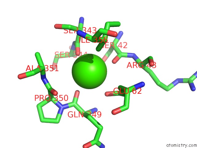
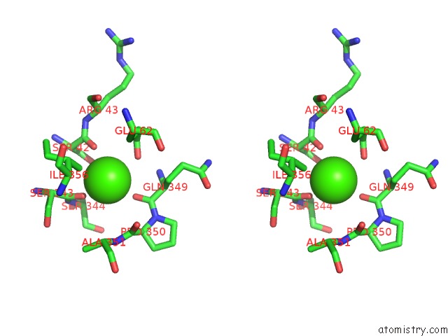
Calcium binding site 1 out of 3 in 4ufc
Go back to
Calcium binding site 1 out
of 3 in the Crystal Structure of the GH95 Enzyme BACOVA_03438
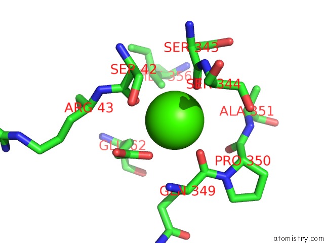
Mono view
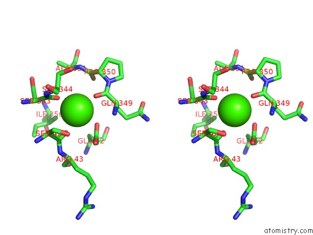
Stereo pair view
Mono view
Stereo pair view
A full contact list of Calcium with other atoms in the Ca binding
site number 1 of Crystal Structure of the GH95 Enzyme BACOVA_03438 within 5.0Å range:
|
Calcium binding site 2 out of 3 in 4ufc
Go back to
Calcium binding site 2 out
of 3 in the Crystal Structure of the GH95 Enzyme BACOVA_03438
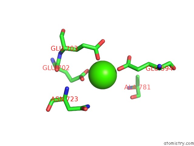
Mono view
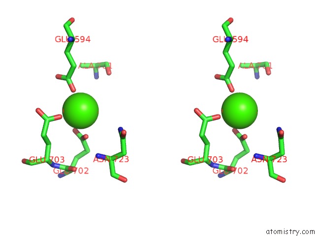
Stereo pair view
Mono view
Stereo pair view
A full contact list of Calcium with other atoms in the Ca binding
site number 2 of Crystal Structure of the GH95 Enzyme BACOVA_03438 within 5.0Å range:
|
Calcium binding site 3 out of 3 in 4ufc
Go back to
Calcium binding site 3 out
of 3 in the Crystal Structure of the GH95 Enzyme BACOVA_03438
Mono view
Stereo pair view
Mono view
Stereo pair view
A full contact list of Calcium with other atoms in the Ca binding
site number 3 of Crystal Structure of the GH95 Enzyme BACOVA_03438 within 5.0Å range:
|
Reference:
A.Rogowski,
J.A.Briggs,
J.C.Mortimer,
T.Tryfona,
N.Terrapon,
E.C.Lowe,
A.Basle,
C.Morland,
A.M.Day,
H.Zheng,
T.E.Rogers,
P.Thompson,
A.R.Hawkins,
M.P.Yadav,
B.Henrissat,
E.C.Martens,
P.Dupree,
H.J.Gilbert,
D.N.Bolam.
Glycan Complexity Dictates Microbial Resource Allocation in the Large Intestine. Nat.Commun. V. 6 7481 2015.
ISSN: ISSN 2041-1723
PubMed: 26112186
DOI: 10.1038/NCOMMS8481
Page generated: Wed Jul 9 02:18:03 2025
ISSN: ISSN 2041-1723
PubMed: 26112186
DOI: 10.1038/NCOMMS8481
Last articles
Cl in 5GUKCl in 5GTZ
Cl in 5GU6
Cl in 5GTI
Cl in 5GTH
Cl in 5GTD
Cl in 5GT3
Cl in 5GT0
Cl in 5GTC
Cl in 5GRQ