Calcium »
PDB 4uj7-4w9y »
4usl »
Calcium in PDB 4usl: The X-Ray Structure of Calcium Bound Human Sorcin
Protein crystallography data
The structure of The X-Ray Structure of Calcium Bound Human Sorcin, PDB code: 4usl
was solved by
A.Ilari,
A.Fiorillo,
G.Colotti,
with X-Ray Crystallography technique. A brief refinement statistics is given in the table below:
| Resolution Low / High (Å) | 37.34 / 1.65 |
| Space group | C 2 2 21 |
| Cell size a, b, c (Å), α, β, γ (°) | 52.369, 111.580, 60.479, 90.00, 90.00, 90.00 |
| R / Rfree (%) | 19.187 / 22.135 |
Calcium Binding Sites:
The binding sites of Calcium atom in the The X-Ray Structure of Calcium Bound Human Sorcin
(pdb code 4usl). This binding sites where shown within
5.0 Angstroms radius around Calcium atom.
In total 3 binding sites of Calcium where determined in the The X-Ray Structure of Calcium Bound Human Sorcin, PDB code: 4usl:
Jump to Calcium binding site number: 1; 2; 3;
In total 3 binding sites of Calcium where determined in the The X-Ray Structure of Calcium Bound Human Sorcin, PDB code: 4usl:
Jump to Calcium binding site number: 1; 2; 3;
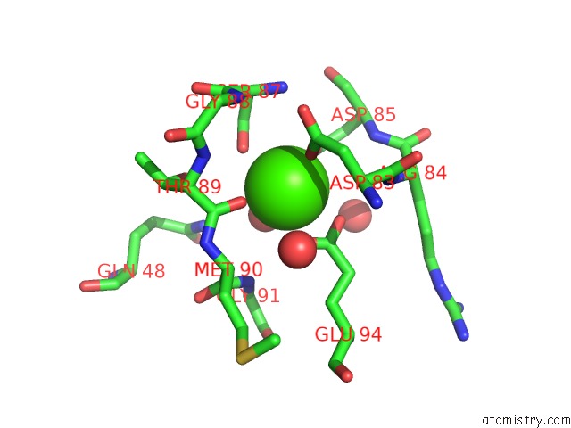
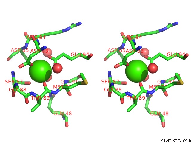
Calcium binding site 1 out of 3 in 4usl
Go back to
Calcium binding site 1 out
of 3 in the The X-Ray Structure of Calcium Bound Human Sorcin
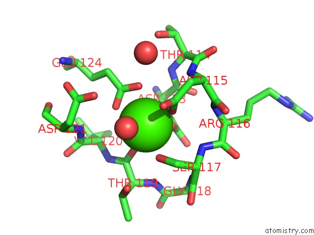
Mono view
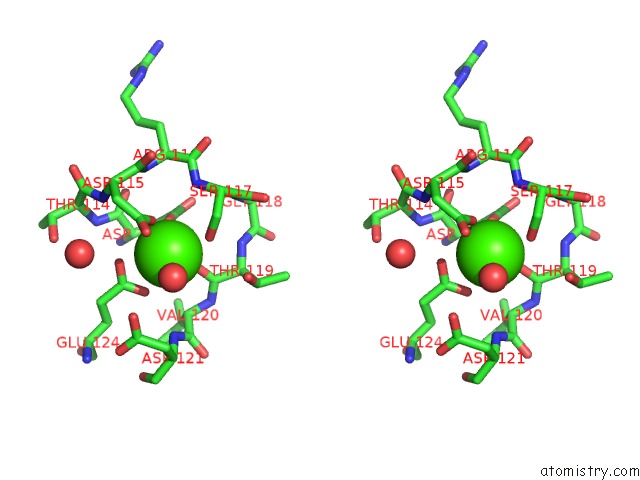
Stereo pair view
Mono view
Stereo pair view
A full contact list of Calcium with other atoms in the Ca binding
site number 1 of The X-Ray Structure of Calcium Bound Human Sorcin within 5.0Å range:
|
Calcium binding site 2 out of 3 in 4usl
Go back to
Calcium binding site 2 out
of 3 in the The X-Ray Structure of Calcium Bound Human Sorcin
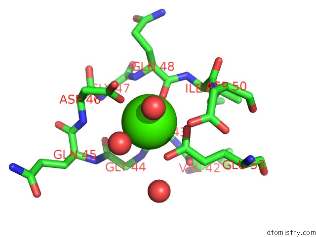
Mono view
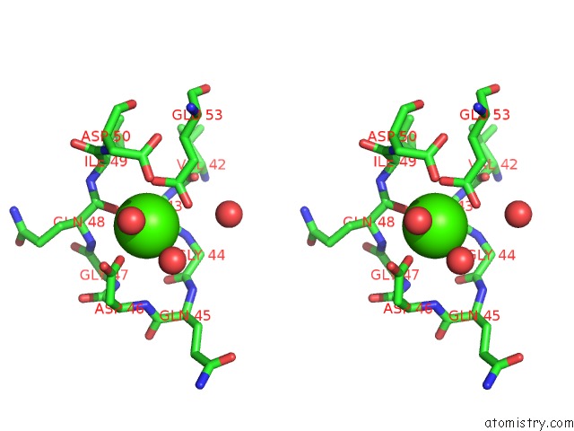
Stereo pair view
Mono view
Stereo pair view
A full contact list of Calcium with other atoms in the Ca binding
site number 2 of The X-Ray Structure of Calcium Bound Human Sorcin within 5.0Å range:
|
Calcium binding site 3 out of 3 in 4usl
Go back to
Calcium binding site 3 out
of 3 in the The X-Ray Structure of Calcium Bound Human Sorcin
Mono view
Stereo pair view
Mono view
Stereo pair view
A full contact list of Calcium with other atoms in the Ca binding
site number 3 of The X-Ray Structure of Calcium Bound Human Sorcin within 5.0Å range:
|
Reference:
A.Ilari,
A.Fiorillo,
E.Poser,
V.S.Lalioti,
G.N.Sundell,
Y.Ivarsson,
I.Genovese,
G.Colotti.
Structural Basis of Sorcin-Mediated Calcium-Dependent Signal Transduction. Sci.Rep. V. 5 16828 2015.
ISSN: ISSN 2045-2322
PubMed: 26577048
DOI: 10.1038/SREP16828
Page generated: Sun Jul 14 13:50:14 2024
ISSN: ISSN 2045-2322
PubMed: 26577048
DOI: 10.1038/SREP16828
Last articles
Zn in 9J0NZn in 9J0O
Zn in 9J0P
Zn in 9FJX
Zn in 9EKB
Zn in 9C0F
Zn in 9CAH
Zn in 9CH0
Zn in 9CH3
Zn in 9CH1