Calcium »
PDB 4uj7-4w9y »
4ut5 »
Calcium in PDB 4ut5: Crystal Structure of the Lecb Lectin From Pseudomonas Aeruginosa Strain PA7 in Complex with Lewis A Tetrasaccharide
Protein crystallography data
The structure of Crystal Structure of the Lecb Lectin From Pseudomonas Aeruginosa Strain PA7 in Complex with Lewis A Tetrasaccharide, PDB code: 4ut5
was solved by
A.M.Boukerb,
A.Decor,
R.Tabaroni,
A.Varrot,
S.Debentzmann,
S.Vidal,
A.Imberty,
B.Cournoyer,
with X-Ray Crystallography technique. A brief refinement statistics is given in the table below:
| Resolution Low / High (Å) | 42.23 / 1.75 |
| Space group | P 1 21 1 |
| Cell size a, b, c (Å), α, β, γ (°) | 52.790, 70.370, 54.660, 90.00, 90.50, 90.00 |
| R / Rfree (%) | 14.752 / 18.345 |
Calcium Binding Sites:
The binding sites of Calcium atom in the Crystal Structure of the Lecb Lectin From Pseudomonas Aeruginosa Strain PA7 in Complex with Lewis A Tetrasaccharide
(pdb code 4ut5). This binding sites where shown within
5.0 Angstroms radius around Calcium atom.
In total 8 binding sites of Calcium where determined in the Crystal Structure of the Lecb Lectin From Pseudomonas Aeruginosa Strain PA7 in Complex with Lewis A Tetrasaccharide, PDB code: 4ut5:
Jump to Calcium binding site number: 1; 2; 3; 4; 5; 6; 7; 8;
In total 8 binding sites of Calcium where determined in the Crystal Structure of the Lecb Lectin From Pseudomonas Aeruginosa Strain PA7 in Complex with Lewis A Tetrasaccharide, PDB code: 4ut5:
Jump to Calcium binding site number: 1; 2; 3; 4; 5; 6; 7; 8;
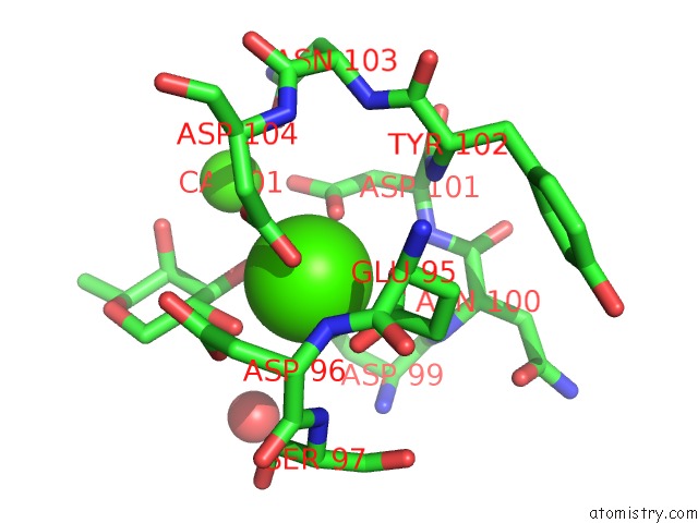
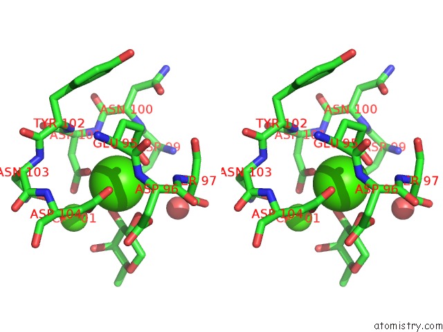
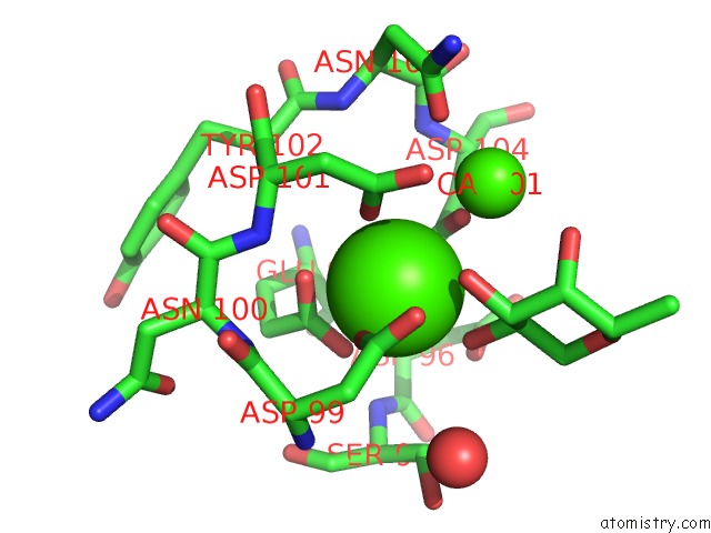
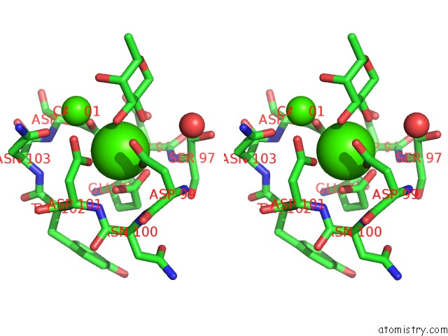
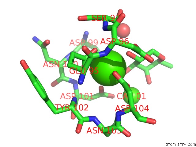
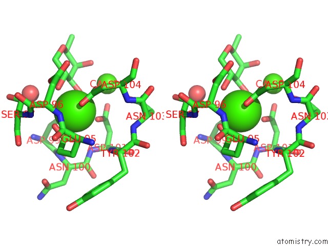
Calcium binding site 1 out of 8 in 4ut5
Go back to
Calcium binding site 1 out
of 8 in the Crystal Structure of the Lecb Lectin From Pseudomonas Aeruginosa Strain PA7 in Complex with Lewis A Tetrasaccharide
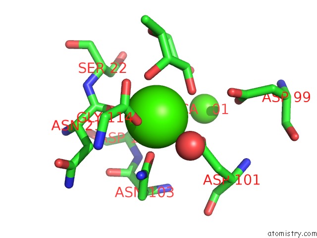
Mono view
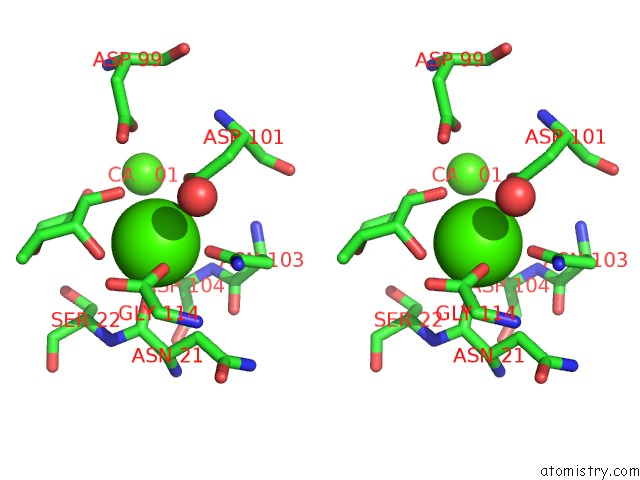
Stereo pair view
Mono view
Stereo pair view
A full contact list of Calcium with other atoms in the Ca binding
site number 1 of Crystal Structure of the Lecb Lectin From Pseudomonas Aeruginosa Strain PA7 in Complex with Lewis A Tetrasaccharide within 5.0Å range:
|
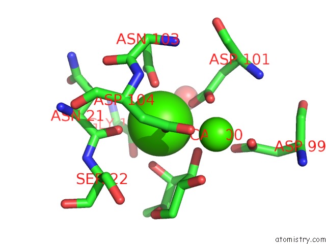
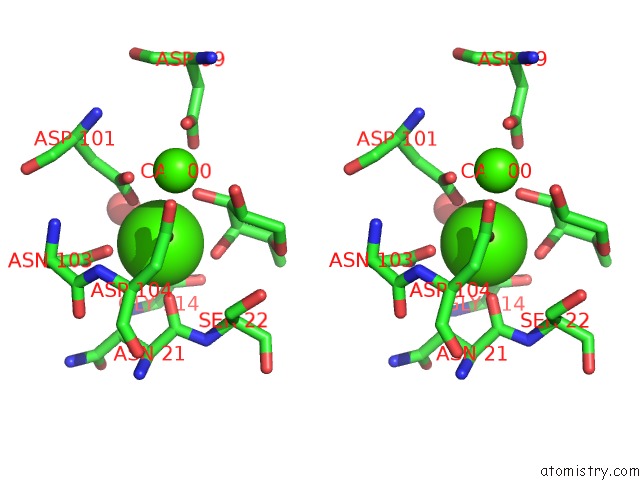
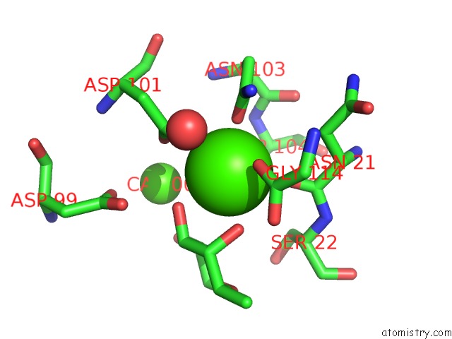
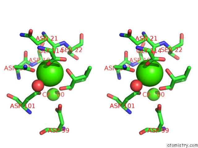
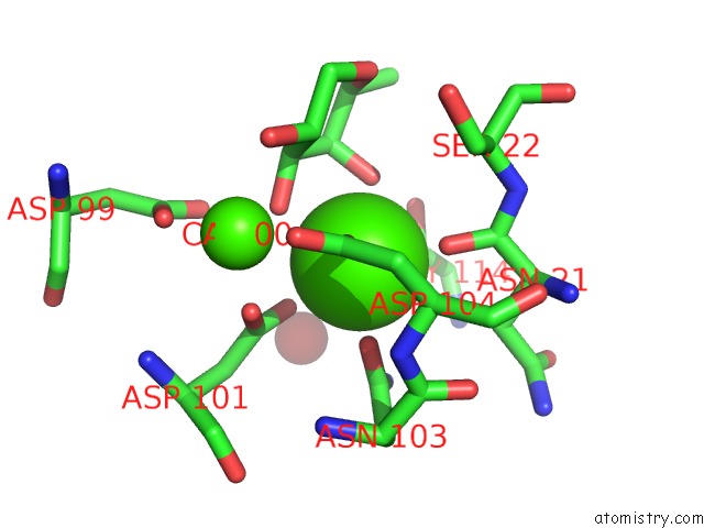
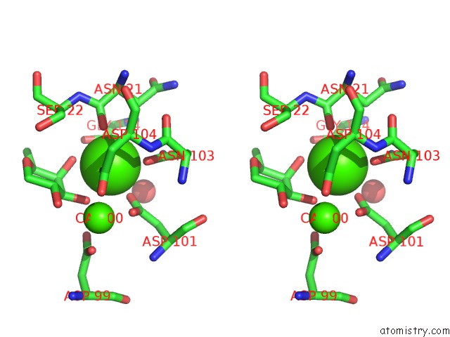
Calcium binding site 2 out of 8 in 4ut5
Go back to
Calcium binding site 2 out
of 8 in the Crystal Structure of the Lecb Lectin From Pseudomonas Aeruginosa Strain PA7 in Complex with Lewis A Tetrasaccharide
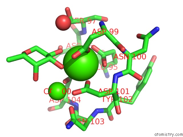
Mono view
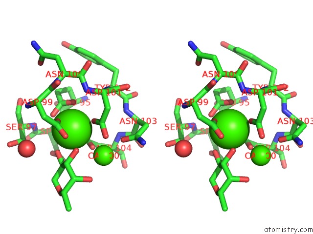
Stereo pair view
Mono view
Stereo pair view
A full contact list of Calcium with other atoms in the Ca binding
site number 2 of Crystal Structure of the Lecb Lectin From Pseudomonas Aeruginosa Strain PA7 in Complex with Lewis A Tetrasaccharide within 5.0Å range:
|
Calcium binding site 3 out of 8 in 4ut5
Go back to
Calcium binding site 3 out
of 8 in the Crystal Structure of the Lecb Lectin From Pseudomonas Aeruginosa Strain PA7 in Complex with Lewis A Tetrasaccharide
Mono view
Stereo pair view
Mono view
Stereo pair view
A full contact list of Calcium with other atoms in the Ca binding
site number 3 of Crystal Structure of the Lecb Lectin From Pseudomonas Aeruginosa Strain PA7 in Complex with Lewis A Tetrasaccharide within 5.0Å range:
|
Calcium binding site 4 out of 8 in 4ut5
Go back to
Calcium binding site 4 out
of 8 in the Crystal Structure of the Lecb Lectin From Pseudomonas Aeruginosa Strain PA7 in Complex with Lewis A Tetrasaccharide
Mono view
Stereo pair view
Mono view
Stereo pair view
A full contact list of Calcium with other atoms in the Ca binding
site number 4 of Crystal Structure of the Lecb Lectin From Pseudomonas Aeruginosa Strain PA7 in Complex with Lewis A Tetrasaccharide within 5.0Å range:
|
Calcium binding site 5 out of 8 in 4ut5
Go back to
Calcium binding site 5 out
of 8 in the Crystal Structure of the Lecb Lectin From Pseudomonas Aeruginosa Strain PA7 in Complex with Lewis A Tetrasaccharide
Mono view
Stereo pair view
Mono view
Stereo pair view
A full contact list of Calcium with other atoms in the Ca binding
site number 5 of Crystal Structure of the Lecb Lectin From Pseudomonas Aeruginosa Strain PA7 in Complex with Lewis A Tetrasaccharide within 5.0Å range:
|
Calcium binding site 6 out of 8 in 4ut5
Go back to
Calcium binding site 6 out
of 8 in the Crystal Structure of the Lecb Lectin From Pseudomonas Aeruginosa Strain PA7 in Complex with Lewis A Tetrasaccharide
Mono view
Stereo pair view
Mono view
Stereo pair view
A full contact list of Calcium with other atoms in the Ca binding
site number 6 of Crystal Structure of the Lecb Lectin From Pseudomonas Aeruginosa Strain PA7 in Complex with Lewis A Tetrasaccharide within 5.0Å range:
|
Calcium binding site 7 out of 8 in 4ut5
Go back to
Calcium binding site 7 out
of 8 in the Crystal Structure of the Lecb Lectin From Pseudomonas Aeruginosa Strain PA7 in Complex with Lewis A Tetrasaccharide
Mono view
Stereo pair view
Mono view
Stereo pair view
A full contact list of Calcium with other atoms in the Ca binding
site number 7 of Crystal Structure of the Lecb Lectin From Pseudomonas Aeruginosa Strain PA7 in Complex with Lewis A Tetrasaccharide within 5.0Å range:
|
Calcium binding site 8 out of 8 in 4ut5
Go back to
Calcium binding site 8 out
of 8 in the Crystal Structure of the Lecb Lectin From Pseudomonas Aeruginosa Strain PA7 in Complex with Lewis A Tetrasaccharide
Mono view
Stereo pair view
Mono view
Stereo pair view
A full contact list of Calcium with other atoms in the Ca binding
site number 8 of Crystal Structure of the Lecb Lectin From Pseudomonas Aeruginosa Strain PA7 in Complex with Lewis A Tetrasaccharide within 5.0Å range:
|
Reference:
A.M.Boukerb,
A.Decor,
S.Ribun,
R.Tabaroni,
A.Rousset,
L.Commin,
S.Buff,
A.Doleans-Jordheim,
S.Vidal,
A.Varrot,
A.Imberty,
B.Cournoyer.
Genomic Rearrangements and Functional Diversification of Leca and Lecb Lectin-Coding Regions Impacting the Efficacy of Glycomimetics Directed Against Pseudomonas Aeruginosa. Front.Microbiol. V. 7 811 2016.
ISSN: ISSN 1664-302X
PubMed: 27303392
DOI: 10.3389/FMICB.2016.00811
Page generated: Wed Jul 9 02:25:05 2025
ISSN: ISSN 1664-302X
PubMed: 27303392
DOI: 10.3389/FMICB.2016.00811
Last articles
F in 4G5PF in 4G5J
F in 4G3G
F in 4G3F
F in 4G2I
F in 4G2H
F in 4G31
F in 4G1W
F in 4G16
F in 4FXY