Calcium »
PDB 4uj7-4w9y »
4v2x »
Calcium in PDB 4v2x: High Resolution Structure of the Full Length Tri-Modular Endo-Beta-1,4-Glucanase B (CEL5B) From Bacillus Halodurans
Protein crystallography data
The structure of High Resolution Structure of the Full Length Tri-Modular Endo-Beta-1,4-Glucanase B (CEL5B) From Bacillus Halodurans, PDB code: 4v2x
was solved by
I.Venditto,
H.Santos,
L.M.A.Ferreira,
K.Sakka,
C.M.G.A.Fontes,
S.Najmudin,
with X-Ray Crystallography technique. A brief refinement statistics is given in the table below:
| Resolution Low / High (Å) | 51.27 / 1.64 |
| Space group | P 21 21 2 |
| Cell size a, b, c (Å), α, β, γ (°) | 74.220, 141.840, 50.820, 90.00, 90.00, 90.00 |
| R / Rfree (%) | 15.43 / 18.094 |
Other elements in 4v2x:
The structure of High Resolution Structure of the Full Length Tri-Modular Endo-Beta-1,4-Glucanase B (CEL5B) From Bacillus Halodurans also contains other interesting chemical elements:
| Arsenic | (As) | 1 atom |
Calcium Binding Sites:
The binding sites of Calcium atom in the High Resolution Structure of the Full Length Tri-Modular Endo-Beta-1,4-Glucanase B (CEL5B) From Bacillus Halodurans
(pdb code 4v2x). This binding sites where shown within
5.0 Angstroms radius around Calcium atom.
In total 2 binding sites of Calcium where determined in the High Resolution Structure of the Full Length Tri-Modular Endo-Beta-1,4-Glucanase B (CEL5B) From Bacillus Halodurans, PDB code: 4v2x:
Jump to Calcium binding site number: 1; 2;
In total 2 binding sites of Calcium where determined in the High Resolution Structure of the Full Length Tri-Modular Endo-Beta-1,4-Glucanase B (CEL5B) From Bacillus Halodurans, PDB code: 4v2x:
Jump to Calcium binding site number: 1; 2;
Calcium binding site 1 out of 2 in 4v2x
Go back to
Calcium binding site 1 out
of 2 in the High Resolution Structure of the Full Length Tri-Modular Endo-Beta-1,4-Glucanase B (CEL5B) From Bacillus Halodurans
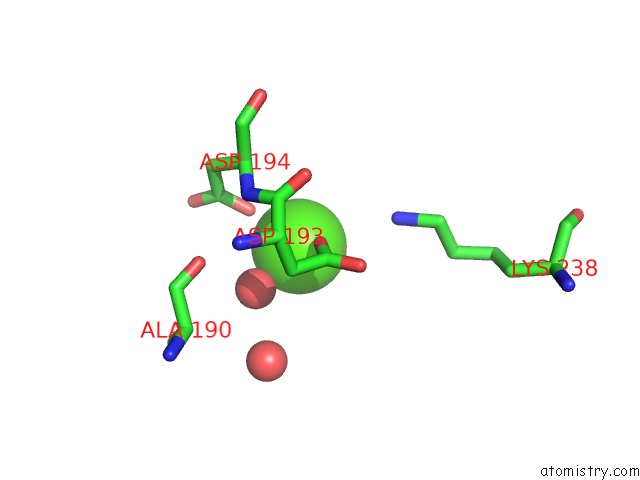
Mono view
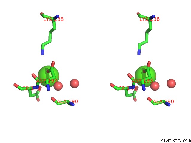
Stereo pair view
Mono view
Stereo pair view
A full contact list of Calcium with other atoms in the Ca binding
site number 1 of High Resolution Structure of the Full Length Tri-Modular Endo-Beta-1,4-Glucanase B (CEL5B) From Bacillus Halodurans within 5.0Å range:
|
Calcium binding site 2 out of 2 in 4v2x
Go back to
Calcium binding site 2 out
of 2 in the High Resolution Structure of the Full Length Tri-Modular Endo-Beta-1,4-Glucanase B (CEL5B) From Bacillus Halodurans
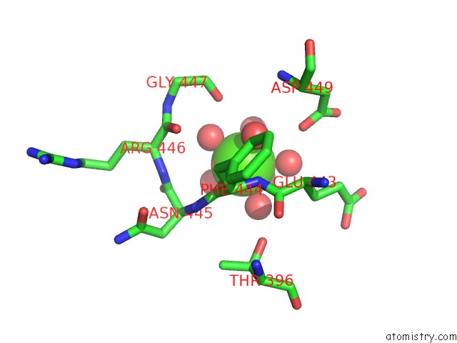
Mono view
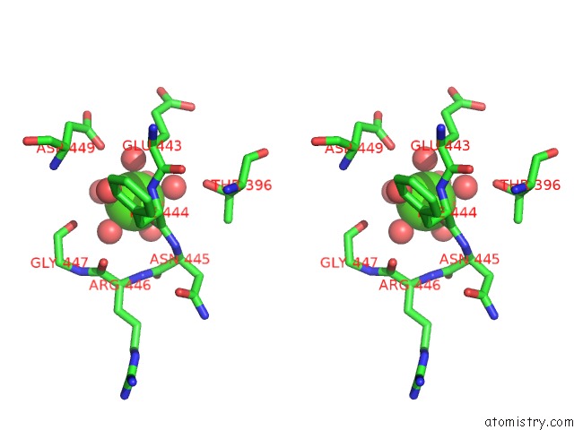
Stereo pair view
Mono view
Stereo pair view
A full contact list of Calcium with other atoms in the Ca binding
site number 2 of High Resolution Structure of the Full Length Tri-Modular Endo-Beta-1,4-Glucanase B (CEL5B) From Bacillus Halodurans within 5.0Å range:
|
Reference:
I.Venditto,
S.Najmudin,
A.S.Luis,
L.M.A.Ferreira,
K.Sakka,
H.J.Gilbert,
C.M.G.A.Fontes.
Family 46 Carbohydrate-Binding Modules Contribute to the Enzymatic Hydrolysis of Xyloglucan and Beta-1,3-1,4-Glucans Through Distinct Mechanisms To Be Published.
Page generated: Sun Jul 14 13:57:16 2024
Last articles
Zn in 9J0NZn in 9J0O
Zn in 9J0P
Zn in 9FJX
Zn in 9EKB
Zn in 9C0F
Zn in 9CAH
Zn in 9CH0
Zn in 9CH3
Zn in 9CH1