Calcium »
PDB 4wa3-4wn0 »
4wa4 »
Calcium in PDB 4wa4: The Crystal Structure of Neuraminidase From A H3N8 Influenza Virus Isolated From New England Harbor Seals in Complex with Oseltamivir Carboxylate
Protein crystallography data
The structure of The Crystal Structure of Neuraminidase From A H3N8 Influenza Virus Isolated From New England Harbor Seals in Complex with Oseltamivir Carboxylate, PDB code: 4wa4
was solved by
H.Yang,
J.M.Villanueva,
L.V.Gubareva,
J.Stevens,
with X-Ray Crystallography technique. A brief refinement statistics is given in the table below:
| Resolution Low / High (Å) | 37.98 / 1.95 |
| Space group | I 4 |
| Cell size a, b, c (Å), α, β, γ (°) | 90.666, 90.666, 108.354, 90.00, 90.00, 90.00 |
| R / Rfree (%) | 13.9 / 16.7 |
Calcium Binding Sites:
The binding sites of Calcium atom in the The Crystal Structure of Neuraminidase From A H3N8 Influenza Virus Isolated From New England Harbor Seals in Complex with Oseltamivir Carboxylate
(pdb code 4wa4). This binding sites where shown within
5.0 Angstroms radius around Calcium atom.
In total only one binding site of Calcium was determined in the The Crystal Structure of Neuraminidase From A H3N8 Influenza Virus Isolated From New England Harbor Seals in Complex with Oseltamivir Carboxylate, PDB code: 4wa4:
In total only one binding site of Calcium was determined in the The Crystal Structure of Neuraminidase From A H3N8 Influenza Virus Isolated From New England Harbor Seals in Complex with Oseltamivir Carboxylate, PDB code: 4wa4:
Calcium binding site 1 out of 1 in 4wa4
Go back to
Calcium binding site 1 out
of 1 in the The Crystal Structure of Neuraminidase From A H3N8 Influenza Virus Isolated From New England Harbor Seals in Complex with Oseltamivir Carboxylate
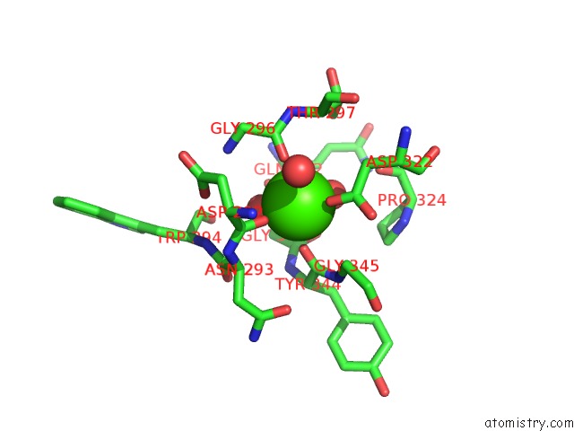
Mono view
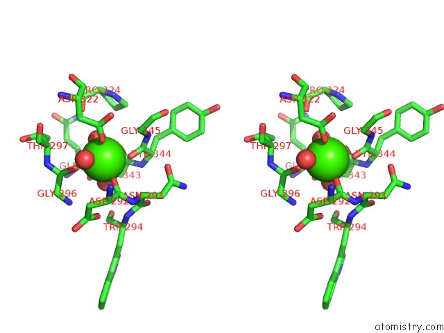
Stereo pair view
Mono view
Stereo pair view
A full contact list of Calcium with other atoms in the Ca binding
site number 1 of The Crystal Structure of Neuraminidase From A H3N8 Influenza Virus Isolated From New England Harbor Seals in Complex with Oseltamivir Carboxylate within 5.0Å range:
|
Reference:
H.Yang,
H.T.Nguyen,
P.J.Carney,
Z.Guo,
J.C.Chang,
J.Jones,
C.T.Davis,
J.M.Villanueva,
L.V.Gubareva,
J.Stevens.
Structural and Functional Analysis of Surface Proteins From An A(H3N8) Influenza Virus Isolated From New England Harbor Seals. J.Virol. 2014.
ISSN: ESSN 1098-5514
PubMed: 25540377
DOI: 10.1128/JVI.02723-14
Page generated: Sun Jul 14 14:03:33 2024
ISSN: ESSN 1098-5514
PubMed: 25540377
DOI: 10.1128/JVI.02723-14
Last articles
Zn in 9J0NZn in 9J0O
Zn in 9J0P
Zn in 9FJX
Zn in 9EKB
Zn in 9C0F
Zn in 9CAH
Zn in 9CH0
Zn in 9CH3
Zn in 9CH1