Calcium »
PDB 4wa3-4wn0 »
4wib »
Calcium in PDB 4wib: Crystal Structure of Magnesium Transporter Mgte
Protein crystallography data
The structure of Crystal Structure of Magnesium Transporter Mgte, PDB code: 4wib
was solved by
H.Takeda,
M.Hattori,
T.Nishizawa,
K.Yamashita,
S.T.A.Shah,
M.Caffrey,
A.D.Maturana,
R.Ishitani,
O.Nureki,
with X-Ray Crystallography technique. A brief refinement statistics is given in the table below:
| Resolution Low / High (Å) | 43.08 / 3.20 |
| Space group | P 21 21 21 |
| Cell size a, b, c (Å), α, β, γ (°) | 64.120, 70.380, 103.340, 90.00, 90.00, 90.00 |
| R / Rfree (%) | 24.1 / 26.8 |
Calcium Binding Sites:
The binding sites of Calcium atom in the Crystal Structure of Magnesium Transporter Mgte
(pdb code 4wib). This binding sites where shown within
5.0 Angstroms radius around Calcium atom.
In total 2 binding sites of Calcium where determined in the Crystal Structure of Magnesium Transporter Mgte, PDB code: 4wib:
Jump to Calcium binding site number: 1; 2;
In total 2 binding sites of Calcium where determined in the Crystal Structure of Magnesium Transporter Mgte, PDB code: 4wib:
Jump to Calcium binding site number: 1; 2;
Calcium binding site 1 out of 2 in 4wib
Go back to
Calcium binding site 1 out
of 2 in the Crystal Structure of Magnesium Transporter Mgte
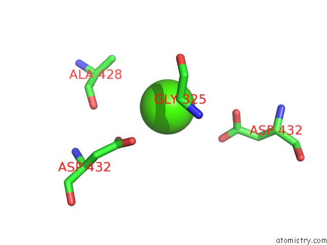
Mono view
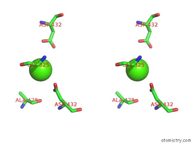
Stereo pair view
Mono view
Stereo pair view
A full contact list of Calcium with other atoms in the Ca binding
site number 1 of Crystal Structure of Magnesium Transporter Mgte within 5.0Å range:
|
Calcium binding site 2 out of 2 in 4wib
Go back to
Calcium binding site 2 out
of 2 in the Crystal Structure of Magnesium Transporter Mgte
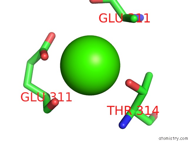
Mono view
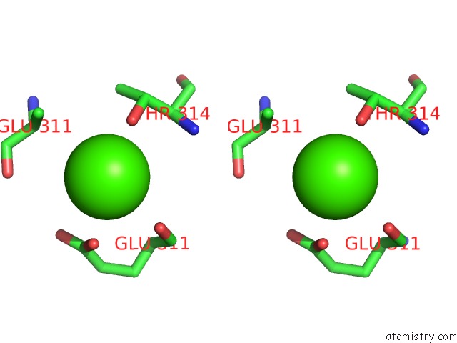
Stereo pair view
Mono view
Stereo pair view
A full contact list of Calcium with other atoms in the Ca binding
site number 2 of Crystal Structure of Magnesium Transporter Mgte within 5.0Å range:
|
Reference:
H.Takeda,
M.Hattori,
T.Nishizawa,
K.Yamashita,
S.T.Shah,
M.Caffrey,
A.D.Maturana,
R.Ishitani,
O.Nureki.
Structural Basis For Ion Selectivity Revealed By High-Resolution Crystal Structure of Mg(2+) Channel Mgte Nat Commun V. 5 5374 2014.
ISSN: ESSN 2041-1723
PubMed: 25367295
DOI: 10.1038/NCOMMS6374
Page generated: Sun Jul 14 14:06:46 2024
ISSN: ESSN 2041-1723
PubMed: 25367295
DOI: 10.1038/NCOMMS6374
Last articles
Zn in 9J0NZn in 9J0O
Zn in 9J0P
Zn in 9FJX
Zn in 9EKB
Zn in 9C0F
Zn in 9CAH
Zn in 9CH0
Zn in 9CH3
Zn in 9CH1