Calcium in PDB 4wis: Crystal Structure of the Lipid Scramblase NHTMEM16 in Crystal Form 1
Protein crystallography data
The structure of Crystal Structure of the Lipid Scramblase NHTMEM16 in Crystal Form 1, PDB code: 4wis
was solved by
R.Dutzler,
J.D.Brunner,
N.K.Lim,
S.Schenck,
with X-Ray Crystallography technique. A brief refinement statistics is given in the table below:
Resolution Low / High (Å)
|
14.98 /
3.30
|
Space group
|
P 21 21 21
|
Cell size a, b, c (Å), α, β, γ (°)
|
96.480,
113.690,
235.650,
90.00,
90.00,
90.00
|
R / Rfree (%)
|
23.8 /
28.5
|
Calcium Binding Sites:
The binding sites of Calcium atom in the Crystal Structure of the Lipid Scramblase NHTMEM16 in Crystal Form 1
(pdb code 4wis). This binding sites where shown within
5.0 Angstroms radius around Calcium atom.
In total 4 binding sites of Calcium where determined in the
Crystal Structure of the Lipid Scramblase NHTMEM16 in Crystal Form 1, PDB code: 4wis:
Jump to Calcium binding site number:
1;
2;
3;
4;
Calcium binding site 1 out
of 4 in 4wis
Go back to
Calcium Binding Sites List in 4wis
Calcium binding site 1 out
of 4 in the Crystal Structure of the Lipid Scramblase NHTMEM16 in Crystal Form 1
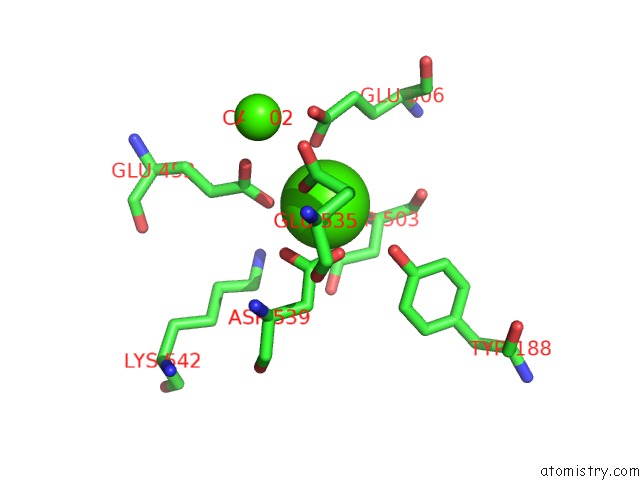
Mono view
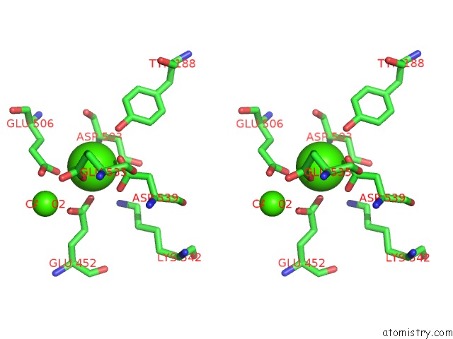
Stereo pair view
|
A full contact list of Calcium with other atoms in the Ca binding
site number 1 of Crystal Structure of the Lipid Scramblase NHTMEM16 in Crystal Form 1 within 5.0Å range:
probe
|
atom
|
residue
|
distance (Å)
|
B
|
Occ
|
A:Ca801
b:96.7
occ:1.00
|
OE2
|
A:GLU535
|
2.5
|
1.0
|
1.0
|
OE1
|
A:GLU452
|
2.5
|
0.6
|
1.0
|
OD2
|
A:ASP539
|
2.5
|
0.9
|
1.0
|
OD1
|
A:ASP539
|
2.6
|
97.8
|
1.0
|
OE2
|
A:GLU506
|
2.7
|
0.9
|
1.0
|
OD1
|
A:ASP503
|
2.7
|
0.4
|
1.0
|
CG
|
A:ASP539
|
2.9
|
0.8
|
1.0
|
CG
|
A:ASP503
|
3.3
|
0.8
|
1.0
|
CD
|
A:GLU452
|
3.4
|
0.0
|
1.0
|
CD
|
A:GLU506
|
3.4
|
0.9
|
1.0
|
OH
|
A:TYR188
|
3.4
|
90.8
|
1.0
|
CD
|
A:GLU535
|
3.5
|
0.3
|
1.0
|
OE2
|
A:GLU452
|
3.6
|
0.7
|
1.0
|
CB
|
A:ASP503
|
3.7
|
0.4
|
1.0
|
CA
|
A:ASP503
|
3.8
|
0.8
|
1.0
|
OE1
|
A:GLU506
|
3.8
|
0.7
|
1.0
|
CG
|
A:GLU535
|
4.1
|
0.8
|
1.0
|
OD2
|
A:ASP503
|
4.2
|
0.4
|
1.0
|
O
|
A:ASP503
|
4.3
|
0.2
|
1.0
|
CB
|
A:ASP539
|
4.4
|
89.1
|
1.0
|
CG
|
A:GLU506
|
4.5
|
0.6
|
1.0
|
CB
|
A:GLU506
|
4.5
|
0.1
|
1.0
|
OE1
|
A:GLU535
|
4.5
|
97.3
|
1.0
|
CZ
|
A:TYR188
|
4.5
|
0.9
|
1.0
|
NZ
|
A:LYS542
|
4.6
|
0.4
|
1.0
|
C
|
A:ASP503
|
4.6
|
0.5
|
1.0
|
CG
|
A:GLU452
|
4.8
|
0.3
|
1.0
|
N
|
A:ASP503
|
4.9
|
0.9
|
1.0
|
CA
|
A:CA802
|
4.9
|
0.7
|
1.0
|
CE
|
A:LYS542
|
5.0
|
93.6
|
1.0
|
|
Calcium binding site 2 out
of 4 in 4wis
Go back to
Calcium Binding Sites List in 4wis
Calcium binding site 2 out
of 4 in the Crystal Structure of the Lipid Scramblase NHTMEM16 in Crystal Form 1
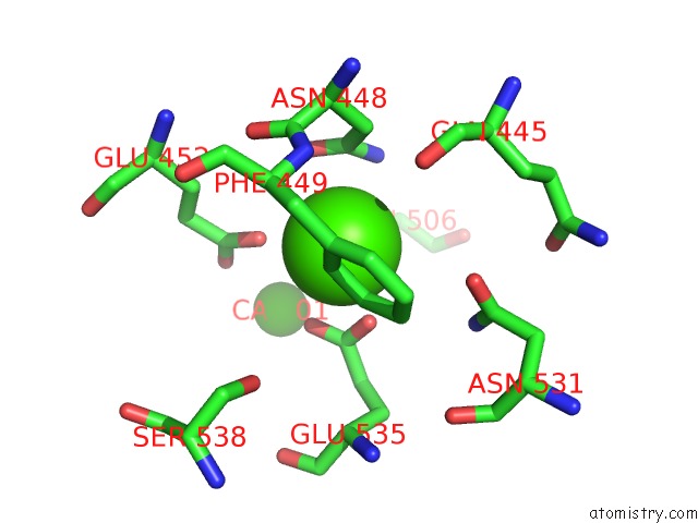
Mono view
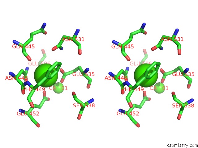
Stereo pair view
|
A full contact list of Calcium with other atoms in the Ca binding
site number 2 of Crystal Structure of the Lipid Scramblase NHTMEM16 in Crystal Form 1 within 5.0Å range:
probe
|
atom
|
residue
|
distance (Å)
|
B
|
Occ
|
A:Ca802
b:0.7
occ:1.00
|
OE1
|
A:GLU535
|
2.5
|
97.3
|
1.0
|
OE1
|
A:GLU506
|
2.6
|
0.7
|
1.0
|
OE2
|
A:GLU452
|
2.9
|
0.7
|
1.0
|
CG
|
A:ASN448
|
3.2
|
0.1
|
1.0
|
OE2
|
A:GLU535
|
3.2
|
1.0
|
1.0
|
CD
|
A:GLU535
|
3.2
|
0.3
|
1.0
|
OD1
|
A:ASN448
|
3.3
|
0.0
|
1.0
|
CD1
|
A:PHE449
|
3.4
|
0.7
|
1.0
|
CE1
|
A:PHE449
|
3.4
|
0.4
|
1.0
|
ND2
|
A:ASN448
|
3.4
|
0.8
|
1.0
|
CD
|
A:GLU506
|
3.7
|
0.9
|
1.0
|
CB
|
A:ASN448
|
3.7
|
0.5
|
1.0
|
OE2
|
A:GLU506
|
4.1
|
0.9
|
1.0
|
CD
|
A:GLU452
|
4.2
|
0.0
|
1.0
|
N
|
A:PHE449
|
4.2
|
0.2
|
1.0
|
C
|
A:ASN448
|
4.2
|
0.3
|
1.0
|
OD1
|
A:ASN531
|
4.4
|
0.3
|
1.0
|
O
|
A:ASN448
|
4.5
|
0.6
|
1.0
|
CA
|
A:ASN448
|
4.6
|
0.9
|
1.0
|
CG
|
A:PHE449
|
4.6
|
0.4
|
1.0
|
CZ
|
A:PHE449
|
4.6
|
0.6
|
1.0
|
CA
|
A:PHE449
|
4.6
|
0.9
|
1.0
|
ND2
|
A:ASN531
|
4.7
|
0.1
|
1.0
|
CG
|
A:GLU535
|
4.7
|
0.8
|
1.0
|
O
|
A:GLN445
|
4.7
|
0.4
|
1.0
|
OE1
|
A:GLU452
|
4.9
|
0.6
|
1.0
|
CB
|
A:GLU452
|
4.9
|
0.4
|
1.0
|
CG
|
A:GLU506
|
4.9
|
0.6
|
1.0
|
CA
|
A:CA801
|
4.9
|
96.7
|
1.0
|
OG
|
A:SER538
|
5.0
|
0.8
|
1.0
|
CG
|
A:ASN531
|
5.0
|
95.5
|
1.0
|
|
Calcium binding site 3 out
of 4 in 4wis
Go back to
Calcium Binding Sites List in 4wis
Calcium binding site 3 out
of 4 in the Crystal Structure of the Lipid Scramblase NHTMEM16 in Crystal Form 1
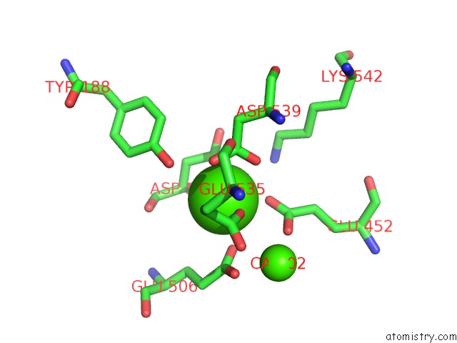
Mono view
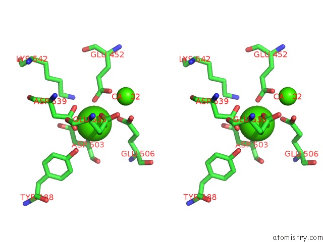
Stereo pair view
|
A full contact list of Calcium with other atoms in the Ca binding
site number 3 of Crystal Structure of the Lipid Scramblase NHTMEM16 in Crystal Form 1 within 5.0Å range:
probe
|
atom
|
residue
|
distance (Å)
|
B
|
Occ
|
B:Ca801
b:0.8
occ:1.00
|
OE2
|
B:GLU535
|
2.5
|
0.6
|
1.0
|
OD2
|
B:ASP539
|
2.5
|
0.7
|
1.0
|
OE1
|
B:GLU452
|
2.6
|
0.5
|
1.0
|
OD1
|
B:ASP503
|
2.7
|
0.1
|
1.0
|
OD1
|
B:ASP539
|
2.7
|
0.6
|
1.0
|
OE2
|
B:GLU506
|
2.7
|
0.7
|
1.0
|
CG
|
B:ASP539
|
2.9
|
0.6
|
1.0
|
CG
|
B:ASP503
|
3.3
|
0.6
|
1.0
|
OH
|
B:TYR188
|
3.4
|
1.0
|
1.0
|
CD
|
B:GLU506
|
3.5
|
1.0
|
1.0
|
CD
|
B:GLU452
|
3.5
|
0.7
|
1.0
|
CD
|
B:GLU535
|
3.5
|
0.7
|
1.0
|
OE2
|
B:GLU452
|
3.6
|
0.4
|
1.0
|
CB
|
B:ASP503
|
3.7
|
0.2
|
1.0
|
CA
|
B:ASP503
|
3.7
|
0.8
|
1.0
|
OE1
|
B:GLU506
|
3.9
|
97.3
|
1.0
|
CG
|
B:GLU535
|
4.1
|
0.9
|
1.0
|
OD2
|
B:ASP503
|
4.1
|
0.3
|
1.0
|
O
|
B:ASP503
|
4.3
|
0.0
|
1.0
|
CB
|
B:ASP539
|
4.4
|
96.1
|
1.0
|
CB
|
B:GLU506
|
4.5
|
92.2
|
1.0
|
CZ
|
B:TYR188
|
4.5
|
0.0
|
1.0
|
C
|
B:ASP503
|
4.5
|
0.2
|
1.0
|
CG
|
B:GLU506
|
4.5
|
0.8
|
1.0
|
OE1
|
B:GLU535
|
4.5
|
0.1
|
1.0
|
NZ
|
B:LYS542
|
4.6
|
0.3
|
1.0
|
CA
|
B:CA802
|
4.7
|
0.6
|
1.0
|
N
|
B:ASP503
|
4.8
|
97.6
|
1.0
|
CG
|
B:GLU452
|
4.9
|
0.2
|
1.0
|
|
Calcium binding site 4 out
of 4 in 4wis
Go back to
Calcium Binding Sites List in 4wis
Calcium binding site 4 out
of 4 in the Crystal Structure of the Lipid Scramblase NHTMEM16 in Crystal Form 1
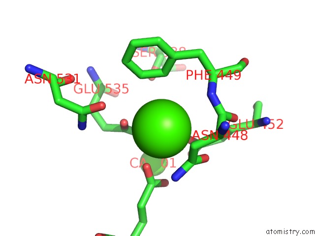
Mono view
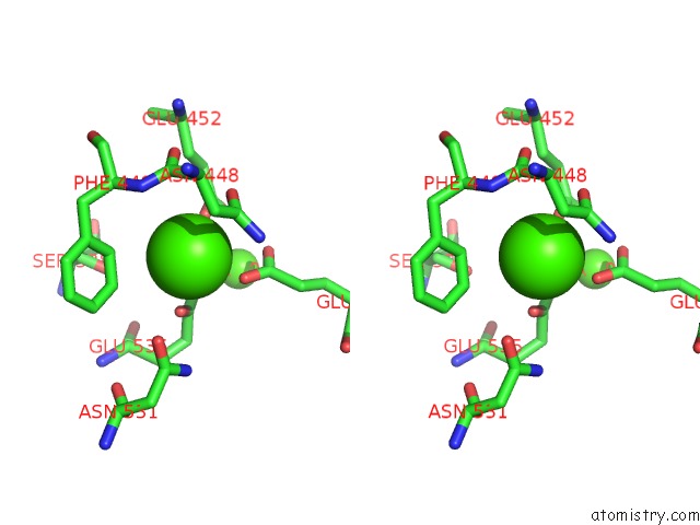
Stereo pair view
|
A full contact list of Calcium with other atoms in the Ca binding
site number 4 of Crystal Structure of the Lipid Scramblase NHTMEM16 in Crystal Form 1 within 5.0Å range:
probe
|
atom
|
residue
|
distance (Å)
|
B
|
Occ
|
B:Ca802
b:0.6
occ:1.00
|
OE1
|
B:GLU535
|
2.5
|
0.1
|
1.0
|
OE1
|
B:GLU506
|
2.5
|
97.3
|
1.0
|
OE2
|
B:GLU452
|
2.6
|
0.4
|
1.0
|
OE2
|
B:GLU535
|
3.0
|
0.6
|
1.0
|
CD
|
B:GLU535
|
3.1
|
0.7
|
1.0
|
OD1
|
B:ASN448
|
3.3
|
1.0
|
1.0
|
CG
|
B:ASN448
|
3.3
|
0.6
|
1.0
|
CD
|
B:GLU506
|
3.5
|
1.0
|
1.0
|
CD1
|
B:PHE449
|
3.6
|
0.9
|
1.0
|
ND2
|
B:ASN448
|
3.6
|
1.0
|
1.0
|
CE1
|
B:PHE449
|
3.6
|
0.1
|
1.0
|
OE2
|
B:GLU506
|
3.8
|
0.7
|
1.0
|
CD
|
B:GLU452
|
3.9
|
0.7
|
1.0
|
CB
|
B:ASN448
|
3.9
|
0.5
|
1.0
|
C
|
B:ASN448
|
4.3
|
1.0
|
1.0
|
N
|
B:PHE449
|
4.4
|
0.5
|
1.0
|
O
|
B:ASN448
|
4.5
|
0.2
|
1.0
|
OE1
|
B:GLU452
|
4.6
|
0.5
|
1.0
|
CG
|
B:GLU535
|
4.6
|
0.9
|
1.0
|
OD1
|
B:ASN531
|
4.6
|
82.6
|
1.0
|
CB
|
B:GLU452
|
4.7
|
0.9
|
1.0
|
CA
|
B:CA801
|
4.7
|
0.8
|
1.0
|
CA
|
B:PHE449
|
4.7
|
0.3
|
1.0
|
CA
|
B:ASN448
|
4.8
|
0.5
|
1.0
|
OG
|
B:SER538
|
4.8
|
0.8
|
1.0
|
CG
|
B:PHE449
|
4.8
|
0.9
|
1.0
|
CZ
|
B:PHE449
|
4.8
|
0.1
|
1.0
|
CG
|
B:GLU506
|
4.9
|
0.8
|
1.0
|
ND2
|
B:ASN531
|
4.9
|
0.1
|
1.0
|
CG
|
B:GLU452
|
4.9
|
0.2
|
1.0
|
|
Reference:
J.D.Brunner,
N.K.Lim,
S.Schenck,
A.Duerst,
R.Dutzler.
X-Ray Structure of A Calcium-Activated TMEM16 Lipid Scramblase. Nature 2014.
ISSN: ESSN 1476-4687
PubMed: 25383531
DOI: 10.1038/NATURE13984
Page generated: Sun Jul 14 14:06:50 2024
|