Calcium »
PDB 4wa3-4wn0 »
4wit »
Calcium in PDB 4wit: TMEM16 Lipid Scramblase in Crystal Form 2
Protein crystallography data
The structure of TMEM16 Lipid Scramblase in Crystal Form 2, PDB code: 4wit
was solved by
R.Dutzler,
J.D.Brunner,
N.K.Lim,
S.Schenck,
with X-Ray Crystallography technique. A brief refinement statistics is given in the table below:
| Resolution Low / High (Å) | 15.00 / 3.40 |
| Space group | P 21 21 21 |
| Cell size a, b, c (Å), α, β, γ (°) | 115.940, 127.240, 180.110, 90.00, 90.00, 90.00 |
| R / Rfree (%) | 24.8 / 29.2 |
Calcium Binding Sites:
The binding sites of Calcium atom in the TMEM16 Lipid Scramblase in Crystal Form 2
(pdb code 4wit). This binding sites where shown within
5.0 Angstroms radius around Calcium atom.
In total 4 binding sites of Calcium where determined in the TMEM16 Lipid Scramblase in Crystal Form 2, PDB code: 4wit:
Jump to Calcium binding site number: 1; 2; 3; 4;
In total 4 binding sites of Calcium where determined in the TMEM16 Lipid Scramblase in Crystal Form 2, PDB code: 4wit:
Jump to Calcium binding site number: 1; 2; 3; 4;
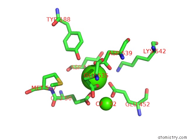
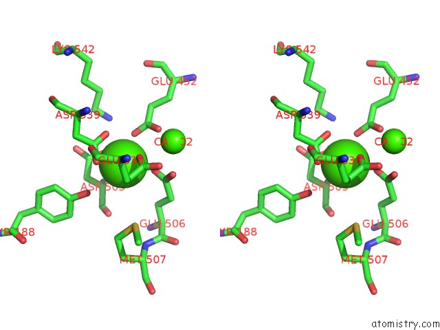
Calcium binding site 1 out of 4 in 4wit
Go back to
Calcium binding site 1 out
of 4 in the TMEM16 Lipid Scramblase in Crystal Form 2
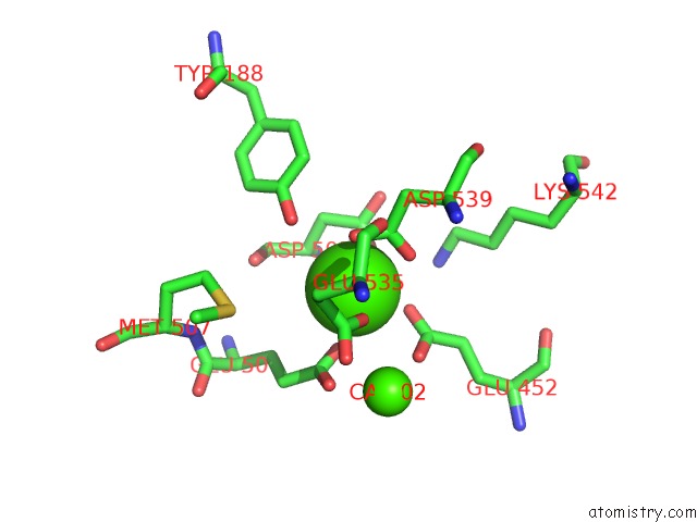
Mono view
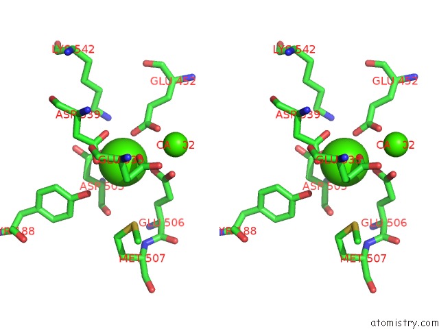
Stereo pair view
Mono view
Stereo pair view
A full contact list of Calcium with other atoms in the Ca binding
site number 1 of TMEM16 Lipid Scramblase in Crystal Form 2 within 5.0Å range:
|
Calcium binding site 2 out of 4 in 4wit
Go back to
Calcium binding site 2 out
of 4 in the TMEM16 Lipid Scramblase in Crystal Form 2
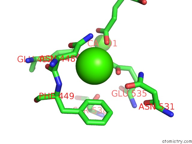
Mono view
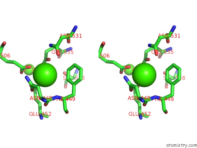
Stereo pair view
Mono view
Stereo pair view
A full contact list of Calcium with other atoms in the Ca binding
site number 2 of TMEM16 Lipid Scramblase in Crystal Form 2 within 5.0Å range:
|
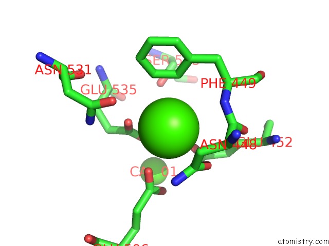
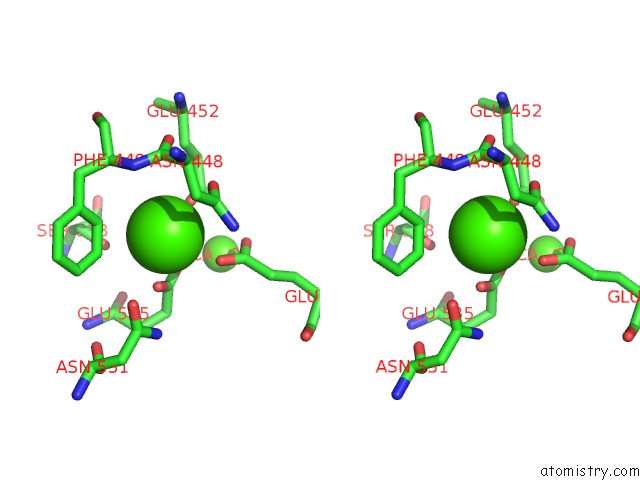
Calcium binding site 3 out of 4 in 4wit
Go back to
Calcium binding site 3 out
of 4 in the TMEM16 Lipid Scramblase in Crystal Form 2
Mono view
Stereo pair view
Mono view
Stereo pair view
A full contact list of Calcium with other atoms in the Ca binding
site number 3 of TMEM16 Lipid Scramblase in Crystal Form 2 within 5.0Å range:
|
Calcium binding site 4 out of 4 in 4wit
Go back to
Calcium binding site 4 out
of 4 in the TMEM16 Lipid Scramblase in Crystal Form 2
Mono view
Stereo pair view
Mono view
Stereo pair view
A full contact list of Calcium with other atoms in the Ca binding
site number 4 of TMEM16 Lipid Scramblase in Crystal Form 2 within 5.0Å range:
|
Reference:
J.D.Brunner,
N.K.Lim,
S.Schenck,
A.Duerst,
R.Dutzler.
X-Ray Structure of A Calcium-Activated TMEM16 Lipid Scramblase. Nature 2014.
ISSN: ESSN 1476-4687
PubMed: 25383531
DOI: 10.1038/NATURE13984
Page generated: Sun Jul 14 14:06:52 2024
ISSN: ESSN 1476-4687
PubMed: 25383531
DOI: 10.1038/NATURE13984
Last articles
Zn in 9J0NZn in 9J0O
Zn in 9J0P
Zn in 9FJX
Zn in 9EKB
Zn in 9C0F
Zn in 9CAH
Zn in 9CH0
Zn in 9CH3
Zn in 9CH1