Calcium »
PDB 4wnb-4x8d »
4wo1 »
Calcium in PDB 4wo1: Crystal Structure of the DAP12 Transmembrane Domain in Lipid Cubic Phase
Protein crystallography data
The structure of Crystal Structure of the DAP12 Transmembrane Domain in Lipid Cubic Phase, PDB code: 4wo1
was solved by
M.J.Call,
M.E.Call,
K.Knoblich,
with X-Ray Crystallography technique. A brief refinement statistics is given in the table below:
| Resolution Low / High (Å) | 33.12 / 2.14 |
| Space group | P 21 21 2 |
| Cell size a, b, c (Å), α, β, γ (°) | 50.797, 43.685, 50.605, 90.00, 90.00, 90.00 |
| R / Rfree (%) | 23.2 / 29.6 |
Calcium Binding Sites:
The binding sites of Calcium atom in the Crystal Structure of the DAP12 Transmembrane Domain in Lipid Cubic Phase
(pdb code 4wo1). This binding sites where shown within
5.0 Angstroms radius around Calcium atom.
In total 2 binding sites of Calcium where determined in the Crystal Structure of the DAP12 Transmembrane Domain in Lipid Cubic Phase, PDB code: 4wo1:
Jump to Calcium binding site number: 1; 2;
In total 2 binding sites of Calcium where determined in the Crystal Structure of the DAP12 Transmembrane Domain in Lipid Cubic Phase, PDB code: 4wo1:
Jump to Calcium binding site number: 1; 2;
Calcium binding site 1 out of 2 in 4wo1
Go back to
Calcium binding site 1 out
of 2 in the Crystal Structure of the DAP12 Transmembrane Domain in Lipid Cubic Phase
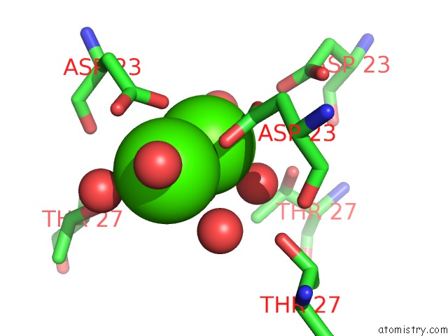
Mono view
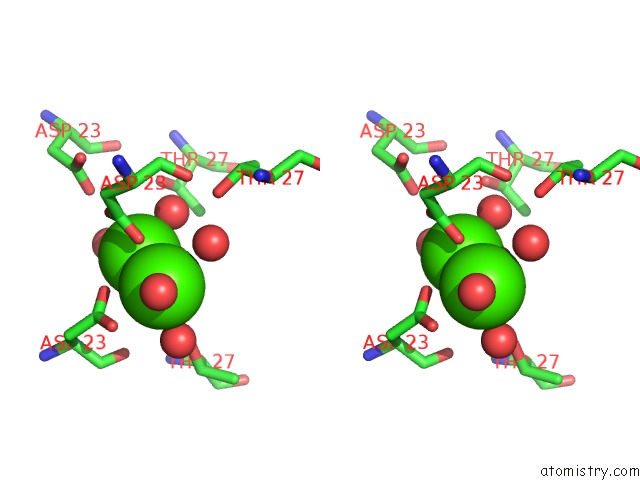
Stereo pair view
Mono view
Stereo pair view
A full contact list of Calcium with other atoms in the Ca binding
site number 1 of Crystal Structure of the DAP12 Transmembrane Domain in Lipid Cubic Phase within 5.0Å range:
|
Calcium binding site 2 out of 2 in 4wo1
Go back to
Calcium binding site 2 out
of 2 in the Crystal Structure of the DAP12 Transmembrane Domain in Lipid Cubic Phase
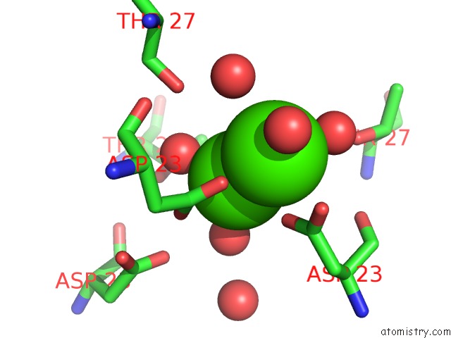
Mono view
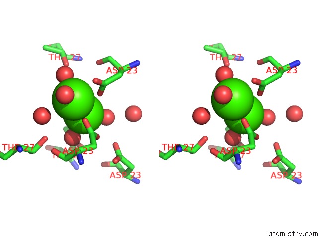
Stereo pair view
Mono view
Stereo pair view
A full contact list of Calcium with other atoms in the Ca binding
site number 2 of Crystal Structure of the DAP12 Transmembrane Domain in Lipid Cubic Phase within 5.0Å range:
|
Reference:
K.Knoblich,
S.Park,
M.Lutfi,
L.Van 't Hag,
C.E.Conn,
S.A.Seabrook,
J.Newman,
P.E.Czabotar,
W.Im,
M.E.Call,
M.J.Call.
Transmembrane Complexes of DAP12 Crystallized in Lipid Membranes Provide Insights Into Control of Oligomerization in Immunoreceptor Assembly. Cell Rep V. 11 1184 2015.
ISSN: ESSN 2211-1247
PubMed: 25981043
DOI: 10.1016/J.CELREP.2015.04.045
Page generated: Wed Jul 9 02:42:28 2025
ISSN: ESSN 2211-1247
PubMed: 25981043
DOI: 10.1016/J.CELREP.2015.04.045
Last articles
Ca in 5L89Ca in 5L7F
Ca in 5L7P
Ca in 5L41
Ca in 5L3U
Ca in 5L73
Ca in 5L79
Ca in 5L30
Ca in 5L2Z
Ca in 5L1I