Calcium »
PDB 4wnb-4x8d »
4wxq »
Calcium in PDB 4wxq: Crystal Structure of the Myocilin Olfactomedin Domain
Protein crystallography data
The structure of Crystal Structure of the Myocilin Olfactomedin Domain, PDB code: 4wxq
was solved by
S.D.Orwig,
K.C.Turnage,
R.K.Donegan,
R.L.Lieberman,
with X-Ray Crystallography technique. A brief refinement statistics is given in the table below:
| Resolution Low / High (Å) | 19.78 / 2.15 |
| Space group | I 2 2 2 |
| Cell size a, b, c (Å), α, β, γ (°) | 68.660, 85.820, 88.440, 90.00, 90.00, 90.00 |
| R / Rfree (%) | 19.3 / 23.1 |
Other elements in 4wxq:
The structure of Crystal Structure of the Myocilin Olfactomedin Domain also contains other interesting chemical elements:
| Sodium | (Na) | 1 atom |
Calcium Binding Sites:
The binding sites of Calcium atom in the Crystal Structure of the Myocilin Olfactomedin Domain
(pdb code 4wxq). This binding sites where shown within
5.0 Angstroms radius around Calcium atom.
In total only one binding site of Calcium was determined in the Crystal Structure of the Myocilin Olfactomedin Domain, PDB code: 4wxq:
In total only one binding site of Calcium was determined in the Crystal Structure of the Myocilin Olfactomedin Domain, PDB code: 4wxq:
Calcium binding site 1 out of 1 in 4wxq
Go back to
Calcium binding site 1 out
of 1 in the Crystal Structure of the Myocilin Olfactomedin Domain
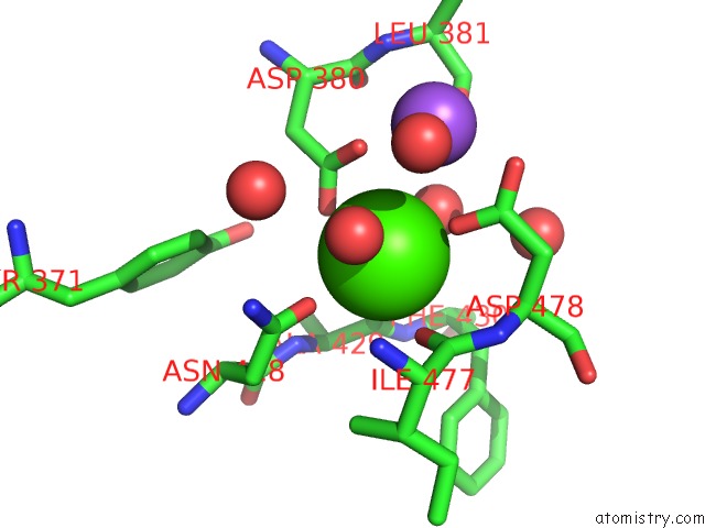
Mono view
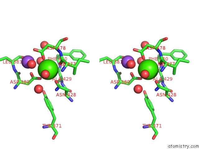
Stereo pair view
Mono view
Stereo pair view
A full contact list of Calcium with other atoms in the Ca binding
site number 1 of Crystal Structure of the Myocilin Olfactomedin Domain within 5.0Å range:
|
Reference:
R.K.Donegan,
S.E.Hill,
D.M.Freeman,
E.Nguyen,
S.D.Orwig,
K.C.Turnage,
R.L.Lieberman.
Structural Basis For Misfolding in Myocilin-Associated Glaucoma. Hum.Mol.Genet. V. 24 2111 2015.
ISSN: ESSN 1460-2083
PubMed: 25524706
DOI: 10.1093/HMG/DDU730
Page generated: Sun Jul 14 14:25:01 2024
ISSN: ESSN 1460-2083
PubMed: 25524706
DOI: 10.1093/HMG/DDU730
Last articles
Zn in 9MJ5Zn in 9HNW
Zn in 9G0L
Zn in 9FNE
Zn in 9DZN
Zn in 9E0I
Zn in 9D32
Zn in 9DAK
Zn in 8ZXC
Zn in 8ZUF