Calcium »
PDB 4x8e-4xur »
4xb3 »
Calcium in PDB 4xb3: Structure of Dextran Glucosidase
Enzymatic activity of Structure of Dextran Glucosidase
All present enzymatic activity of Structure of Dextran Glucosidase:
3.2.1.70;
3.2.1.70;
Protein crystallography data
The structure of Structure of Dextran Glucosidase, PDB code: 4xb3
was solved by
M.Kobayashi,
K.Kato,
M.Yao,
with X-Ray Crystallography technique. A brief refinement statistics is given in the table below:
| Resolution Low / High (Å) | 48.11 / 2.09 |
| Space group | P 21 21 21 |
| Cell size a, b, c (Å), α, β, γ (°) | 72.151, 82.550, 103.559, 90.00, 90.00, 90.00 |
| R / Rfree (%) | 17.9 / 21.9 |
Calcium Binding Sites:
The binding sites of Calcium atom in the Structure of Dextran Glucosidase
(pdb code 4xb3). This binding sites where shown within
5.0 Angstroms radius around Calcium atom.
In total 4 binding sites of Calcium where determined in the Structure of Dextran Glucosidase, PDB code: 4xb3:
Jump to Calcium binding site number: 1; 2; 3; 4;
In total 4 binding sites of Calcium where determined in the Structure of Dextran Glucosidase, PDB code: 4xb3:
Jump to Calcium binding site number: 1; 2; 3; 4;
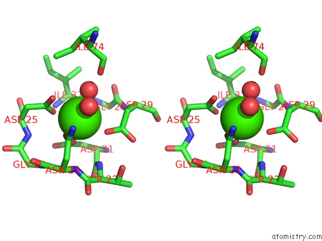
Calcium binding site 1 out of 4 in 4xb3
Go back to
Calcium binding site 1 out
of 4 in the Structure of Dextran Glucosidase

Mono view

Stereo pair view
Mono view
Stereo pair view
A full contact list of Calcium with other atoms in the Ca binding
site number 1 of Structure of Dextran Glucosidase within 5.0Å range:
|
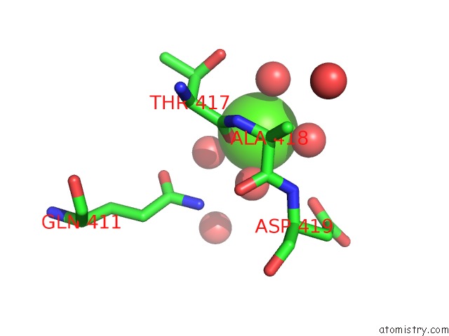
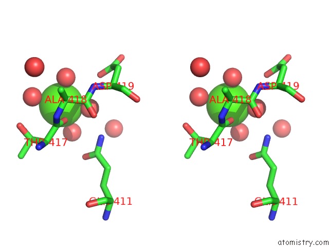
Calcium binding site 2 out of 4 in 4xb3
Go back to
Calcium binding site 2 out
of 4 in the Structure of Dextran Glucosidase

Mono view
Stereo pair view
Mono view
Stereo pair view
A full contact list of Calcium with other atoms in the Ca binding
site number 2 of Structure of Dextran Glucosidase within 5.0Å range:
|
Calcium binding site 3 out of 4 in 4xb3
Go back to
Calcium binding site 3 out
of 4 in the Structure of Dextran Glucosidase
Mono view
Stereo pair view
Mono view
Stereo pair view
A full contact list of Calcium with other atoms in the Ca binding
site number 3 of Structure of Dextran Glucosidase within 5.0Å range:
|
Calcium binding site 4 out of 4 in 4xb3
Go back to
Calcium binding site 4 out
of 4 in the Structure of Dextran Glucosidase

Mono view
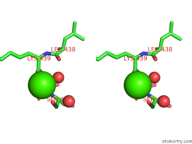
Stereo pair view
Mono view
Stereo pair view
A full contact list of Calcium with other atoms in the Ca binding
site number 4 of Structure of Dextran Glucosidase within 5.0Å range:
|
Reference:
M.Kobayashi,
W.Saburi,
D.Nakatsuka,
H.Hondoh,
K.Kato,
M.Okuyama,
H.Mori,
A.Kimura,
M.Yao.
Structural Insights Into the Catalytic Reaction That Is Involved in the Reorientation of TRP238 at the Substrate-Binding Site in GH13 Dextran Glucosidase Febs Lett. V. 589 484 2015.
ISSN: ISSN 0014-5793
PubMed: 25595454
DOI: 10.1016/J.FEBSLET.2015.01.005
Page generated: Sun Jul 14 14:32:50 2024
ISSN: ISSN 0014-5793
PubMed: 25595454
DOI: 10.1016/J.FEBSLET.2015.01.005
Last articles
Zn in 9J0NZn in 9J0O
Zn in 9J0P
Zn in 9FJX
Zn in 9EKB
Zn in 9C0F
Zn in 9CAH
Zn in 9CH0
Zn in 9CH3
Zn in 9CH1