Calcium »
PDB 4x8e-4xur »
4xhz »
Calcium in PDB 4xhz: Crystal Structure of Human Protocadherin-15 EC8-10
Protein crystallography data
The structure of Crystal Structure of Human Protocadherin-15 EC8-10, PDB code: 4xhz
was solved by
R.Araya-Secchi,
M.Sotomayor,
with X-Ray Crystallography technique. A brief refinement statistics is given in the table below:
| Resolution Low / High (Å) | 136.77 / 2.80 |
| Space group | P 63 2 2 |
| Cell size a, b, c (Å), α, β, γ (°) | 157.929, 157.929, 142.898, 90.00, 90.00, 120.00 |
| R / Rfree (%) | 17 / 20 |
Other elements in 4xhz:
The structure of Crystal Structure of Human Protocadherin-15 EC8-10 also contains other interesting chemical elements:
| Chlorine | (Cl) | 1 atom |
Calcium Binding Sites:
The binding sites of Calcium atom in the Crystal Structure of Human Protocadherin-15 EC8-10
(pdb code 4xhz). This binding sites where shown within
5.0 Angstroms radius around Calcium atom.
In total 3 binding sites of Calcium where determined in the Crystal Structure of Human Protocadherin-15 EC8-10, PDB code: 4xhz:
Jump to Calcium binding site number: 1; 2; 3;
In total 3 binding sites of Calcium where determined in the Crystal Structure of Human Protocadherin-15 EC8-10, PDB code: 4xhz:
Jump to Calcium binding site number: 1; 2; 3;
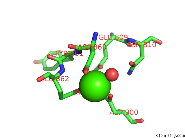
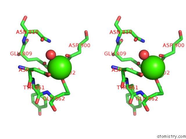
Calcium binding site 1 out of 3 in 4xhz
Go back to
Calcium binding site 1 out
of 3 in the Crystal Structure of Human Protocadherin-15 EC8-10
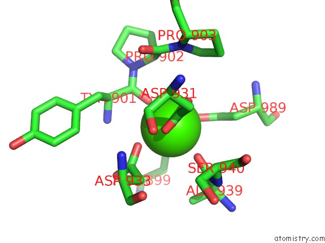
Mono view
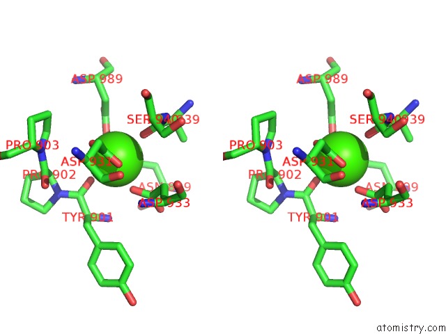
Stereo pair view
Mono view
Stereo pair view
A full contact list of Calcium with other atoms in the Ca binding
site number 1 of Crystal Structure of Human Protocadherin-15 EC8-10 within 5.0Å range:
|
Calcium binding site 2 out of 3 in 4xhz
Go back to
Calcium binding site 2 out
of 3 in the Crystal Structure of Human Protocadherin-15 EC8-10
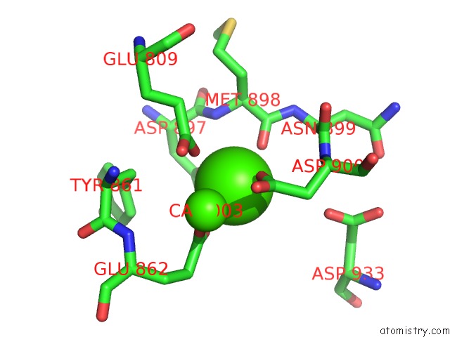
Mono view
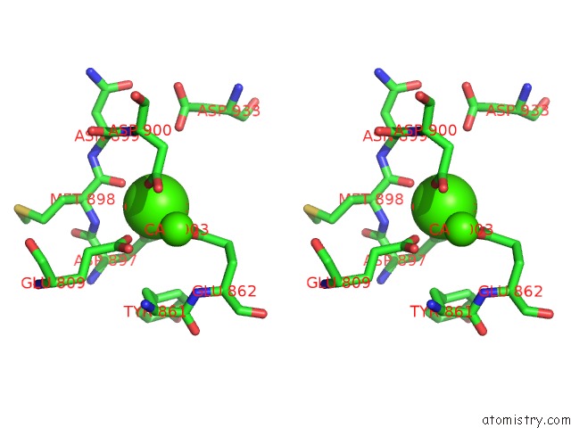
Stereo pair view
Mono view
Stereo pair view
A full contact list of Calcium with other atoms in the Ca binding
site number 2 of Crystal Structure of Human Protocadherin-15 EC8-10 within 5.0Å range:
|
Calcium binding site 3 out of 3 in 4xhz
Go back to
Calcium binding site 3 out
of 3 in the Crystal Structure of Human Protocadherin-15 EC8-10
Mono view
Stereo pair view
Mono view
Stereo pair view
A full contact list of Calcium with other atoms in the Ca binding
site number 3 of Crystal Structure of Human Protocadherin-15 EC8-10 within 5.0Å range:
|
Reference:
R.Araya-Secchi,
B.L.Neel,
M.Sotomayor.
An Elastic Element in the Protocadherin-15 Tip Link of the Inner Ear. Nat Commun V. 7 13458 2016.
ISSN: ESSN 2041-1723
PubMed: 27857071
DOI: 10.1038/NCOMMS13458
Page generated: Wed Jul 9 02:52:28 2025
ISSN: ESSN 2041-1723
PubMed: 27857071
DOI: 10.1038/NCOMMS13458
Last articles
F in 7LCRF in 7LCM
F in 7LCO
F in 7LCK
F in 7LCJ
F in 7LCI
F in 7L9Y
F in 7LCD
F in 7LAY
F in 7LAJ