Calcium »
PDB 4x8e-4xur »
4xuq »
Calcium in PDB 4xuq: Structure of the CBM22-2 Xylan-Binding Domain From Paenibacillus Barcinonensis XYN10C in Complex with Xylotriose
Enzymatic activity of Structure of the CBM22-2 Xylan-Binding Domain From Paenibacillus Barcinonensis XYN10C in Complex with Xylotriose
All present enzymatic activity of Structure of the CBM22-2 Xylan-Binding Domain From Paenibacillus Barcinonensis XYN10C in Complex with Xylotriose:
3.2.1.8;
3.2.1.8;
Protein crystallography data
The structure of Structure of the CBM22-2 Xylan-Binding Domain From Paenibacillus Barcinonensis XYN10C in Complex with Xylotriose, PDB code: 4xuq
was solved by
M.A.Sainz-Polo,
J.Sanz-Aparicio,
with X-Ray Crystallography technique. A brief refinement statistics is given in the table below:
| Resolution Low / High (Å) | 48.42 / 1.95 |
| Space group | P 32 |
| Cell size a, b, c (Å), α, β, γ (°) | 92.455, 92.455, 48.425, 90.00, 90.00, 120.00 |
| R / Rfree (%) | 17.8 / 21.3 |
Calcium Binding Sites:
The binding sites of Calcium atom in the Structure of the CBM22-2 Xylan-Binding Domain From Paenibacillus Barcinonensis XYN10C in Complex with Xylotriose
(pdb code 4xuq). This binding sites where shown within
5.0 Angstroms radius around Calcium atom.
In total 3 binding sites of Calcium where determined in the Structure of the CBM22-2 Xylan-Binding Domain From Paenibacillus Barcinonensis XYN10C in Complex with Xylotriose, PDB code: 4xuq:
Jump to Calcium binding site number: 1; 2; 3;
In total 3 binding sites of Calcium where determined in the Structure of the CBM22-2 Xylan-Binding Domain From Paenibacillus Barcinonensis XYN10C in Complex with Xylotriose, PDB code: 4xuq:
Jump to Calcium binding site number: 1; 2; 3;
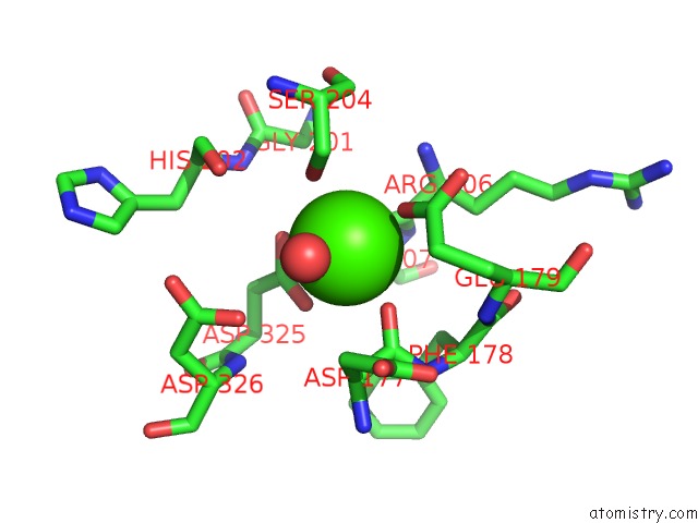
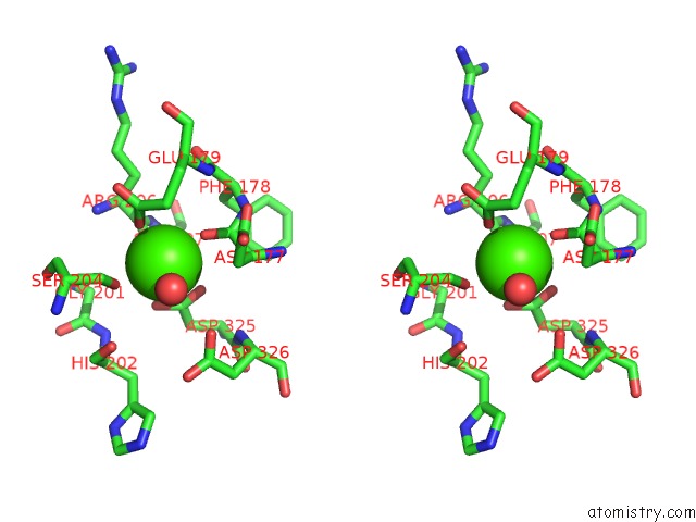
Calcium binding site 1 out of 3 in 4xuq
Go back to
Calcium binding site 1 out
of 3 in the Structure of the CBM22-2 Xylan-Binding Domain From Paenibacillus Barcinonensis XYN10C in Complex with Xylotriose
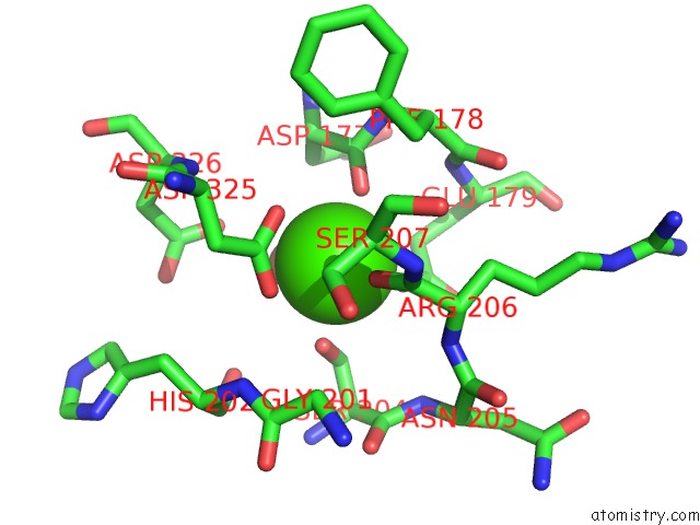
Mono view
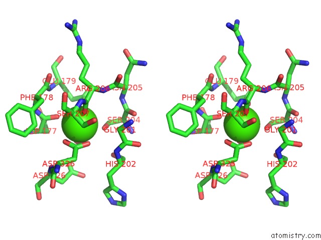
Stereo pair view
Mono view
Stereo pair view
A full contact list of Calcium with other atoms in the Ca binding
site number 1 of Structure of the CBM22-2 Xylan-Binding Domain From Paenibacillus Barcinonensis XYN10C in Complex with Xylotriose within 5.0Å range:
|
Calcium binding site 2 out of 3 in 4xuq
Go back to
Calcium binding site 2 out
of 3 in the Structure of the CBM22-2 Xylan-Binding Domain From Paenibacillus Barcinonensis XYN10C in Complex with Xylotriose
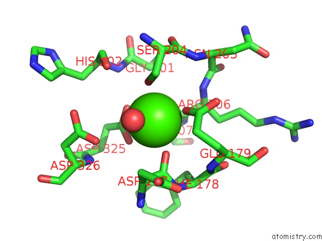
Mono view
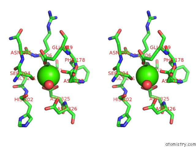
Stereo pair view
Mono view
Stereo pair view
A full contact list of Calcium with other atoms in the Ca binding
site number 2 of Structure of the CBM22-2 Xylan-Binding Domain From Paenibacillus Barcinonensis XYN10C in Complex with Xylotriose within 5.0Å range:
|
Calcium binding site 3 out of 3 in 4xuq
Go back to
Calcium binding site 3 out
of 3 in the Structure of the CBM22-2 Xylan-Binding Domain From Paenibacillus Barcinonensis XYN10C in Complex with Xylotriose
Mono view
Stereo pair view
Mono view
Stereo pair view
A full contact list of Calcium with other atoms in the Ca binding
site number 3 of Structure of the CBM22-2 Xylan-Binding Domain From Paenibacillus Barcinonensis XYN10C in Complex with Xylotriose within 5.0Å range:
|
Reference:
M.A.Sainz-Polo,
B.Gonzalez,
M.Menendez,
F.I.Pastor,
J.Sanz-Aparicio.
Exploring Multimodularity in Plant Cell Wall Deconstruction: Structural and Functional Analysis of XYN10C Containing the CBM22-1-CBM22-2 Tandem. J.Biol.Chem. V. 290 17116 2015.
ISSN: ESSN 1083-351X
PubMed: 26001782
DOI: 10.1074/JBC.M115.659300
Page generated: Sun Jul 14 14:40:59 2024
ISSN: ESSN 1083-351X
PubMed: 26001782
DOI: 10.1074/JBC.M115.659300
Last articles
Zn in 9J0NZn in 9J0O
Zn in 9J0P
Zn in 9FJX
Zn in 9EKB
Zn in 9C0F
Zn in 9CAH
Zn in 9CH0
Zn in 9CH3
Zn in 9CH1