Calcium in PDB 4y8e: PA3825-Eal Ca-Apo Structure
Protein crystallography data
The structure of PA3825-Eal Ca-Apo Structure, PDB code: 4y8e
was solved by
S.Horrell,
D.Bellini,
R.Strange,
A.Wagner,
M.Walsh,
with X-Ray Crystallography technique. A brief refinement statistics is given in the table below:
Resolution Low / High (Å)
|
40.21 /
1.61
|
Space group
|
C 1 2 1
|
Cell size a, b, c (Å), α, β, γ (°)
|
112.440,
59.820,
96.690,
90.00,
116.94,
90.00
|
R / Rfree (%)
|
18.5 /
22.3
|
Calcium Binding Sites:
The binding sites of Calcium atom in the PA3825-Eal Ca-Apo Structure
(pdb code 4y8e). This binding sites where shown within
5.0 Angstroms radius around Calcium atom.
In total 6 binding sites of Calcium where determined in the
PA3825-Eal Ca-Apo Structure, PDB code: 4y8e:
Jump to Calcium binding site number:
1;
2;
3;
4;
5;
6;
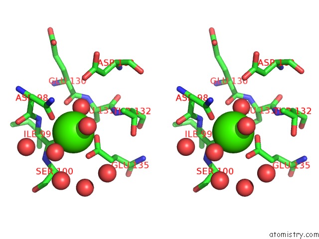
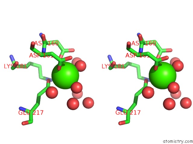
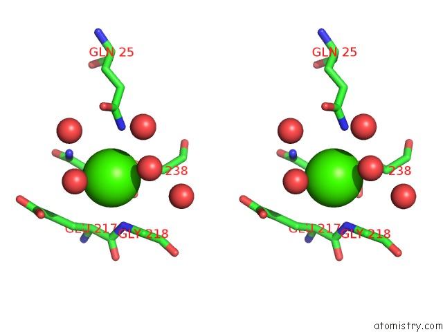
Calcium binding site 1 out
of 6 in 4y8e
Go back to
Calcium Binding Sites List in 4y8e
Calcium binding site 1 out
of 6 in the PA3825-Eal Ca-Apo Structure
 Mono view
 Stereo pair view
|
A full contact list of Calcium with other atoms in the Ca binding
site number 1 of PA3825-Eal Ca-Apo Structure within 5.0Å range:
probe
|
atom
|
residue
|
distance (Å)
|
B
|
Occ
|
A:Ca301
b:21.6
occ:1.00
|
OD1
|
A:ASN98
|
2.3
|
24.1
|
1.0
|
O
|
A:HOH467
|
2.3
|
20.4
|
1.0
|
OE2
|
A:GLU135
|
2.4
|
21.5
|
1.0
|
O
|
A:ILE99
|
2.4
|
19.9
|
1.0
|
O
|
A:HOH535
|
2.5
|
26.6
|
1.0
|
O
|
A:HOH520
|
2.5
|
23.2
|
1.0
|
OE1
|
A:GLU135
|
2.6
|
25.1
|
1.0
|
CD
|
A:GLU135
|
2.8
|
24.0
|
1.0
|
CG
|
A:ASN98
|
3.4
|
22.8
|
1.0
|
C
|
A:ILE99
|
3.4
|
20.7
|
1.0
|
ND2
|
A:ASN98
|
4.0
|
25.3
|
1.0
|
N
|
A:ILE99
|
4.0
|
20.3
|
1.0
|
O
|
A:GLU130
|
4.1
|
19.0
|
1.0
|
N
|
A:SER100
|
4.2
|
20.5
|
1.0
|
O
|
A:HOH497
|
4.2
|
39.8
|
1.0
|
CA
|
A:SER100
|
4.3
|
21.8
|
1.0
|
CG
|
A:GLU135
|
4.3
|
23.2
|
1.0
|
C
|
A:ASN98
|
4.4
|
19.9
|
1.0
|
CA
|
A:ILE99
|
4.4
|
20.5
|
1.0
|
CB
|
A:ASN98
|
4.7
|
20.2
|
1.0
|
CG2
|
A:THR132
|
4.7
|
22.3
|
1.0
|
O
|
A:HOH402
|
4.7
|
40.5
|
1.0
|
N
|
A:THR132
|
4.7
|
21.3
|
1.0
|
O
|
A:HOH445
|
4.7
|
73.9
|
1.0
|
OD2
|
A:ASP160
|
4.8
|
23.1
|
1.0
|
CA
|
A:ASN98
|
4.8
|
18.2
|
1.0
|
CA
|
A:LEU131
|
4.8
|
19.3
|
1.0
|
O
|
A:HOH456
|
4.8
|
27.1
|
1.0
|
O
|
A:ASN98
|
4.9
|
22.8
|
1.0
|
|
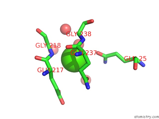
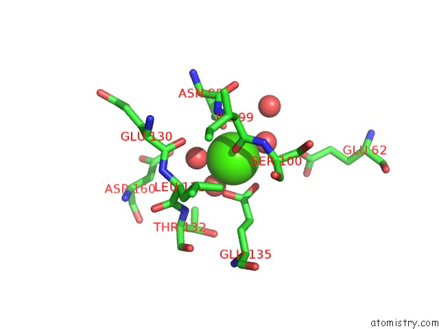
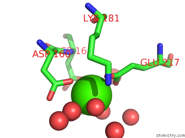
Calcium binding site 2 out
of 6 in 4y8e
Go back to
Calcium Binding Sites List in 4y8e
Calcium binding site 2 out
of 6 in the PA3825-Eal Ca-Apo Structure
 Mono view
 Stereo pair view
|
A full contact list of Calcium with other atoms in the Ca binding
site number 2 of PA3825-Eal Ca-Apo Structure within 5.0Å range:
probe
|
atom
|
residue
|
distance (Å)
|
B
|
Occ
|
A:Ca302
b:29.8
occ:1.00
|
O
|
A:HOH543
|
2.8
|
31.6
|
1.0
|
N
|
A:GLY218
|
3.2
|
16.8
|
1.0
|
OE1
|
A:GLN25
|
3.3
|
35.4
|
1.0
|
OE1
|
A:GLN237
|
3.4
|
31.4
|
1.0
|
CA
|
A:GLY238
|
3.8
|
17.9
|
1.0
|
CG
|
A:GLN237
|
3.8
|
22.3
|
1.0
|
O
|
A:GLN237
|
3.9
|
18.0
|
1.0
|
N
|
A:GLY238
|
3.9
|
16.8
|
1.0
|
C
|
A:GLN237
|
3.9
|
16.3
|
1.0
|
CA
|
A:GLY218
|
4.0
|
16.6
|
1.0
|
CD
|
A:GLN237
|
4.0
|
23.6
|
1.0
|
C
|
A:GLU217
|
4.2
|
16.2
|
1.0
|
CB
|
A:GLU217
|
4.2
|
16.5
|
1.0
|
CD
|
A:GLN25
|
4.2
|
27.0
|
1.0
|
CA
|
A:GLU217
|
4.2
|
16.6
|
1.0
|
NE2
|
A:GLN25
|
4.3
|
26.8
|
1.0
|
O
|
A:HOH462
|
4.3
|
19.6
|
1.0
|
CB
|
A:GLN237
|
4.4
|
21.1
|
1.0
|
O
|
A:HOH552
|
4.6
|
48.4
|
1.0
|
O
|
A:HOH558
|
4.7
|
46.2
|
1.0
|
CG
|
A:GLU217
|
4.8
|
17.8
|
1.0
|
CA
|
A:GLN237
|
4.8
|
17.2
|
1.0
|
OE1
|
A:GLU217
|
4.9
|
24.6
|
1.0
|
|
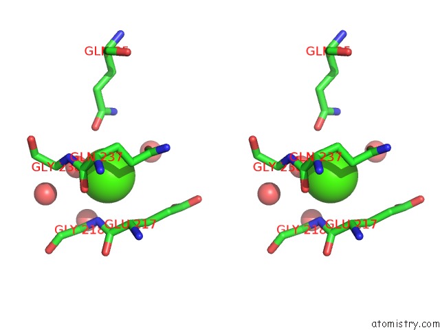
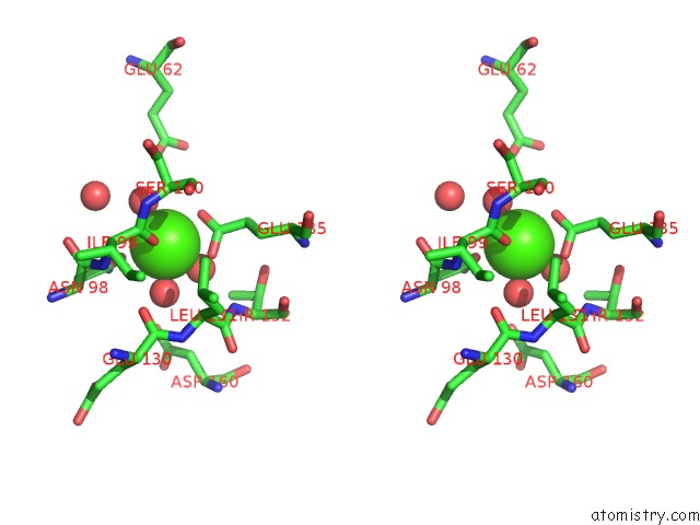
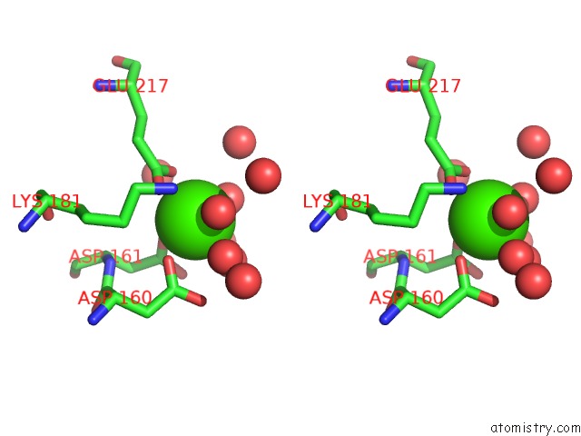
Calcium binding site 3 out
of 6 in 4y8e
Go back to
Calcium Binding Sites List in 4y8e
Calcium binding site 3 out
of 6 in the PA3825-Eal Ca-Apo Structure
 Mono view
 Stereo pair view
|
A full contact list of Calcium with other atoms in the Ca binding
site number 3 of PA3825-Eal Ca-Apo Structure within 5.0Å range:
probe
|
atom
|
residue
|
distance (Å)
|
B
|
Occ
|
A:Ca303
b:23.4
occ:1.00
|
OD1
|
A:ASP161
|
2.3
|
27.5
|
1.0
|
O
|
A:HOH489
|
2.3
|
32.2
|
1.0
|
O
|
A:HOH427
|
2.3
|
26.8
|
1.0
|
OD1
|
A:ASP160
|
2.4
|
22.4
|
1.0
|
OE1
|
A:GLU217
|
2.4
|
24.6
|
1.0
|
O
|
A:HOH514
|
2.4
|
28.8
|
1.0
|
OE2
|
A:GLU217
|
2.5
|
25.1
|
1.0
|
CD
|
A:GLU217
|
2.8
|
22.1
|
1.0
|
CG
|
A:ASP161
|
3.4
|
29.9
|
1.0
|
CG
|
A:ASP160
|
3.5
|
23.1
|
1.0
|
OD2
|
A:ASP160
|
3.7
|
23.1
|
1.0
|
OD2
|
A:ASP161
|
3.8
|
37.9
|
1.0
|
N
|
A:ASP161
|
4.2
|
21.4
|
1.0
|
O
|
A:HOH455
|
4.2
|
27.7
|
1.0
|
O
|
A:HOH431
|
4.2
|
22.6
|
1.0
|
CG
|
A:GLU217
|
4.3
|
17.8
|
1.0
|
NZ
|
A:LYS181
|
4.4
|
23.0
|
1.0
|
CB
|
A:ASP161
|
4.6
|
23.4
|
1.0
|
CA
|
A:ASP161
|
4.7
|
22.0
|
1.0
|
O
|
A:HOH558
|
4.8
|
46.2
|
1.0
|
CB
|
A:ASP160
|
4.8
|
22.0
|
1.0
|
O
|
A:HOH532
|
4.8
|
27.6
|
1.0
|
O
|
A:HOH548
|
4.9
|
50.7
|
1.0
|
C
|
A:ASP160
|
4.9
|
22.0
|
1.0
|
|
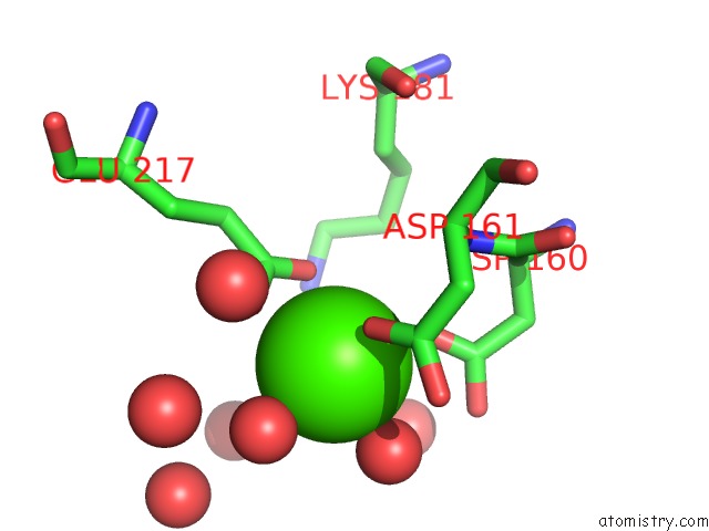
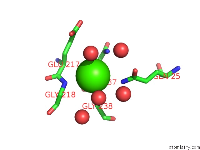
Calcium binding site 4 out
of 6 in 4y8e
Go back to
Calcium Binding Sites List in 4y8e
Calcium binding site 4 out
of 6 in the PA3825-Eal Ca-Apo Structure
 Mono view
 Stereo pair view
|
A full contact list of Calcium with other atoms in the Ca binding
site number 4 of PA3825-Eal Ca-Apo Structure within 5.0Å range:
probe
|
atom
|
residue
|
distance (Å)
|
B
|
Occ
|
B:Ca301
b:25.6
occ:1.00
|
O
|
B:HOH401
|
2.2
|
59.2
|
1.0
|
OD1
|
B:ASN98
|
2.3
|
29.8
|
1.0
|
O
|
B:HOH529
|
2.3
|
29.9
|
1.0
|
O
|
B:ILE99
|
2.4
|
23.4
|
1.0
|
OE2
|
B:GLU135
|
2.4
|
26.6
|
1.0
|
O
|
B:HOH456
|
2.4
|
24.4
|
1.0
|
O
|
B:HOH415
|
2.4
|
31.2
|
1.0
|
OE1
|
B:GLU135
|
2.6
|
26.3
|
1.0
|
CD
|
B:GLU135
|
2.9
|
33.6
|
1.0
|
C
|
B:ILE99
|
3.4
|
25.9
|
1.0
|
CG
|
B:ASN98
|
3.4
|
23.8
|
1.0
|
ND2
|
B:ASN98
|
4.0
|
27.6
|
1.0
|
N
|
B:ILE99
|
4.2
|
22.6
|
1.0
|
N
|
B:SER100
|
4.2
|
26.7
|
1.0
|
CA
|
B:SER100
|
4.3
|
24.2
|
1.0
|
O
|
B:GLU130
|
4.3
|
21.4
|
1.0
|
O
|
B:HOH528
|
4.3
|
47.1
|
1.0
|
CG
|
B:GLU135
|
4.4
|
28.0
|
1.0
|
CA
|
B:ILE99
|
4.4
|
22.7
|
1.0
|
C
|
B:ASN98
|
4.6
|
25.1
|
1.0
|
OE2
|
B:GLU62
|
4.6
|
45.7
|
1.0
|
CB
|
B:ASN98
|
4.7
|
21.1
|
1.0
|
OD2
|
B:ASP160
|
4.7
|
27.7
|
1.0
|
CG2
|
B:THR132
|
4.8
|
24.6
|
1.0
|
N
|
B:THR132
|
4.8
|
22.2
|
1.0
|
CA
|
B:LEU131
|
4.8
|
24.2
|
1.0
|
CA
|
B:ASN98
|
4.9
|
22.2
|
1.0
|
|
Calcium binding site 5 out
of 6 in 4y8e
Go back to
Calcium Binding Sites List in 4y8e
Calcium binding site 5 out
of 6 in the PA3825-Eal Ca-Apo Structure
 Mono view
 Stereo pair view
|
A full contact list of Calcium with other atoms in the Ca binding
site number 5 of PA3825-Eal Ca-Apo Structure within 5.0Å range:
probe
|
atom
|
residue
|
distance (Å)
|
B
|
Occ
|
B:Ca302
b:28.7
occ:1.00
|
O
|
B:HOH546
|
3.1
|
31.2
|
1.0
|
O
|
B:HOH531
|
3.1
|
47.4
|
1.0
|
N
|
B:GLY218
|
3.3
|
18.2
|
1.0
|
NE2
|
B:GLN25
|
3.3
|
27.5
|
1.0
|
NE2
|
B:GLN237
|
3.4
|
26.9
|
1.0
|
CG
|
B:GLN237
|
3.8
|
24.1
|
1.0
|
CA
|
B:GLY238
|
3.8
|
18.0
|
1.0
|
N
|
B:GLY238
|
3.9
|
16.2
|
1.0
|
C
|
B:GLN237
|
3.9
|
18.3
|
1.0
|
O
|
B:GLN237
|
3.9
|
17.9
|
1.0
|
CA
|
B:GLY218
|
4.0
|
19.9
|
1.0
|
CD
|
B:GLN237
|
4.1
|
26.7
|
1.0
|
CB
|
B:GLU217
|
4.1
|
19.6
|
1.0
|
CA
|
B:GLU217
|
4.2
|
17.6
|
1.0
|
C
|
B:GLU217
|
4.2
|
19.3
|
1.0
|
O
|
B:HOH494
|
4.3
|
36.2
|
1.0
|
CD
|
B:GLN25
|
4.3
|
25.6
|
1.0
|
CB
|
B:GLN237
|
4.4
|
22.6
|
1.0
|
OE1
|
B:GLN25
|
4.4
|
28.6
|
1.0
|
O
|
B:HOH493
|
4.4
|
20.3
|
1.0
|
O
|
B:HOH532
|
4.5
|
45.6
|
1.0
|
CG
|
B:GLU217
|
4.8
|
18.9
|
1.0
|
CA
|
B:GLN237
|
4.8
|
18.8
|
1.0
|
OE1
|
B:GLU217
|
4.8
|
31.6
|
1.0
|
|
Calcium binding site 6 out
of 6 in 4y8e
Go back to
Calcium Binding Sites List in 4y8e
Calcium binding site 6 out
of 6 in the PA3825-Eal Ca-Apo Structure
 Mono view
 Stereo pair view
|
A full contact list of Calcium with other atoms in the Ca binding
site number 6 of PA3825-Eal Ca-Apo Structure within 5.0Å range:
probe
|
atom
|
residue
|
distance (Å)
|
B
|
Occ
|
B:Ca303
b:27.4
occ:1.00
|
O
|
B:HOH503
|
2.2
|
45.4
|
1.0
|
O
|
B:HOH487
|
2.3
|
39.0
|
1.0
|
OD1
|
B:ASP161
|
2.4
|
34.0
|
1.0
|
OD1
|
B:ASP160
|
2.4
|
28.3
|
1.0
|
O
|
B:HOH481
|
2.5
|
41.6
|
1.0
|
OE1
|
B:GLU217
|
2.5
|
31.6
|
1.0
|
OE2
|
B:GLU217
|
2.5
|
27.2
|
1.0
|
CD
|
B:GLU217
|
2.9
|
26.4
|
1.0
|
CG
|
B:ASP160
|
3.4
|
28.1
|
1.0
|
CG
|
B:ASP161
|
3.4
|
37.5
|
1.0
|
O
|
B:HOH428
|
3.5
|
28.4
|
1.0
|
OD2
|
B:ASP160
|
3.8
|
27.7
|
1.0
|
OD2
|
B:ASP161
|
3.9
|
38.6
|
1.0
|
O
|
B:HOH410
|
4.0
|
28.0
|
1.0
|
O
|
B:HOH531
|
4.2
|
47.4
|
1.0
|
O
|
B:HOH418
|
4.3
|
44.6
|
1.0
|
N
|
B:ASP161
|
4.3
|
26.3
|
1.0
|
CG
|
B:GLU217
|
4.4
|
18.9
|
1.0
|
NZ
|
B:LYS181
|
4.5
|
23.4
|
1.0
|
O
|
B:HOH522
|
4.7
|
36.0
|
1.0
|
CB
|
B:ASP161
|
4.7
|
31.1
|
1.0
|
CB
|
B:ASP160
|
4.8
|
24.4
|
1.0
|
O
|
B:HOH486
|
4.8
|
34.8
|
1.0
|
CA
|
B:ASP161
|
4.8
|
29.1
|
1.0
|
|
Reference:
D.Bellini,
S.Horrell,
R.Strange,
A.Wagner,
A.Hitchin,
J.S.Webb,
I.Tews,
M.A.Walch.
Structure of PA3825 From P. Aeruginosa Bound to Cyclic Di-Gmp and Pgpg: New Insights For A Potential Three-Metal Catalytic Mechanism of Eal Domains To Be Published.
Page generated: Sun Jul 14 14:50:30 2024
|