Calcium »
PDB 4y9p-4yty »
4yj2 »
Calcium in PDB 4yj2: Crystal Structure of Tubulin Bound to Mi-181
Protein crystallography data
The structure of Crystal Structure of Tubulin Bound to Mi-181, PDB code: 4yj2
was solved by
D.E.Mcnamara,
J.Z.Torres,
T.O.Yeates,
with X-Ray Crystallography technique. A brief refinement statistics is given in the table below:
| Resolution Low / High (Å) | 90.72 / 2.60 |
| Space group | P 21 21 21 |
| Cell size a, b, c (Å), α, β, γ (°) | 104.830, 157.650, 181.030, 90.00, 90.00, 90.00 |
| R / Rfree (%) | 18.8 / 23.1 |
Other elements in 4yj2:
The structure of Crystal Structure of Tubulin Bound to Mi-181 also contains other interesting chemical elements:
| Magnesium | (Mg) | 4 atoms |
Calcium Binding Sites:
The binding sites of Calcium atom in the Crystal Structure of Tubulin Bound to Mi-181
(pdb code 4yj2). This binding sites where shown within
5.0 Angstroms radius around Calcium atom.
In total 3 binding sites of Calcium where determined in the Crystal Structure of Tubulin Bound to Mi-181, PDB code: 4yj2:
Jump to Calcium binding site number: 1; 2; 3;
In total 3 binding sites of Calcium where determined in the Crystal Structure of Tubulin Bound to Mi-181, PDB code: 4yj2:
Jump to Calcium binding site number: 1; 2; 3;
Calcium binding site 1 out of 3 in 4yj2
Go back to
Calcium binding site 1 out
of 3 in the Crystal Structure of Tubulin Bound to Mi-181

Mono view
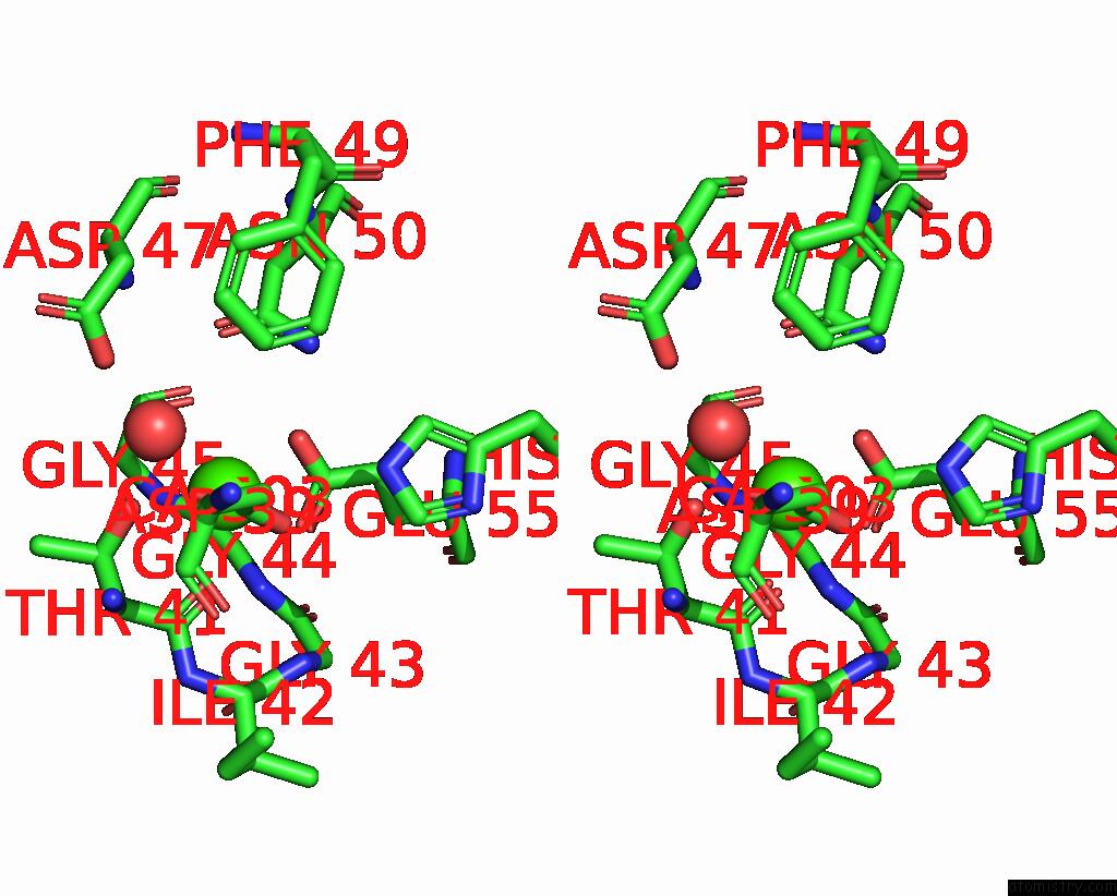
Stereo pair view
Mono view
Stereo pair view
A full contact list of Calcium with other atoms in the Ca binding
site number 1 of Crystal Structure of Tubulin Bound to Mi-181 within 5.0Å range:
|
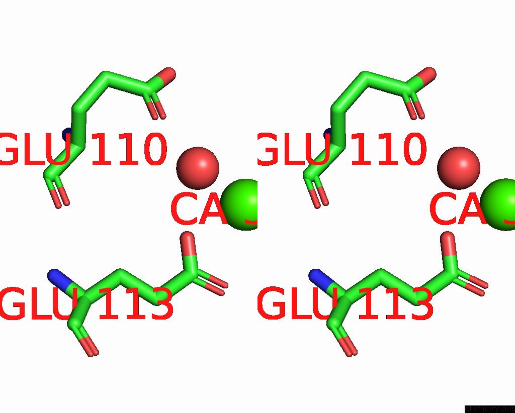
Calcium binding site 2 out of 3 in 4yj2
Go back to
Calcium binding site 2 out
of 3 in the Crystal Structure of Tubulin Bound to Mi-181
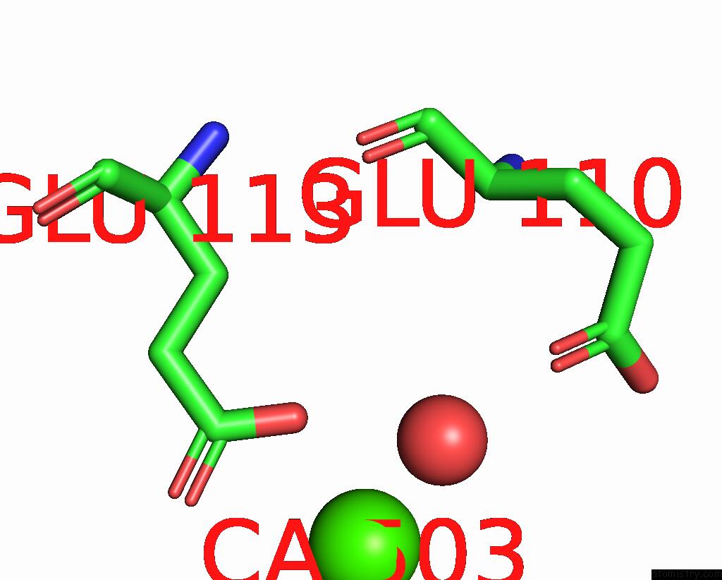
Mono view
Stereo pair view
Mono view
Stereo pair view
A full contact list of Calcium with other atoms in the Ca binding
site number 2 of Crystal Structure of Tubulin Bound to Mi-181 within 5.0Å range:
|
Calcium binding site 3 out of 3 in 4yj2
Go back to
Calcium binding site 3 out
of 3 in the Crystal Structure of Tubulin Bound to Mi-181

Mono view
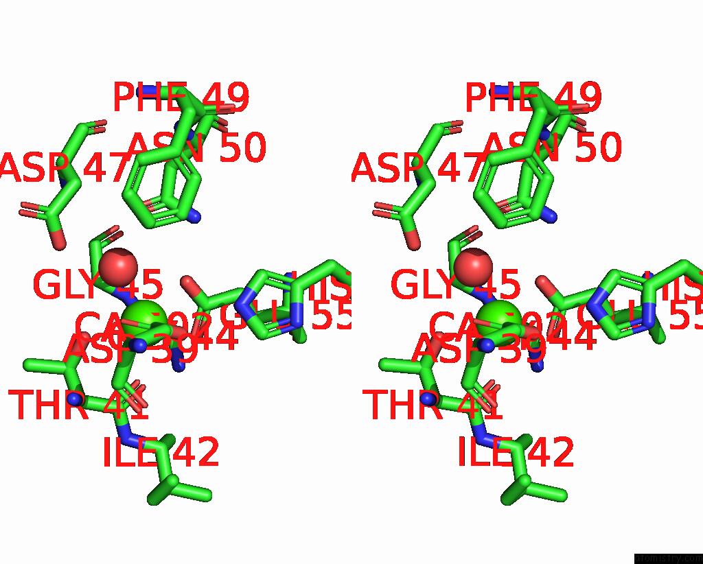
Stereo pair view
Mono view
Stereo pair view
A full contact list of Calcium with other atoms in the Ca binding
site number 3 of Crystal Structure of Tubulin Bound to Mi-181 within 5.0Å range:
|
Reference:
D.E.Mcnamara,
S.Senese,
T.O.Yeates,
J.Z.Torres.
Structures of Potent Anticancer Compounds Bound to Tubulin. Protein Sci. 2015.
ISSN: ESSN 1469-896X
PubMed: 25970265
DOI: 10.1002/PRO.2704
Page generated: Wed Jul 9 03:12:29 2025
ISSN: ESSN 1469-896X
PubMed: 25970265
DOI: 10.1002/PRO.2704
Last articles
Fe in 2YXOFe in 2YRS
Fe in 2YXC
Fe in 2YNM
Fe in 2YVJ
Fe in 2YP1
Fe in 2YU2
Fe in 2YU1
Fe in 2YQB
Fe in 2YOO