Calcium »
PDB 4z72-4zmp »
4zmo »
Calcium in PDB 4zmo: Crystal Structure of Human P-Cadherin (Ss-Dimer K14E)
Protein crystallography data
The structure of Crystal Structure of Human P-Cadherin (Ss-Dimer K14E), PDB code: 4zmo
was solved by
J.M.M.Caaveiro,
S.Kudo,
K.Tsumoto,
with X-Ray Crystallography technique. A brief refinement statistics is given in the table below:
| Resolution Low / High (Å) | 42.43 / 2.48 |
| Space group | C 1 2 1 |
| Cell size a, b, c (Å), α, β, γ (°) | 118.380, 81.230, 48.990, 90.00, 105.79, 90.00 |
| R / Rfree (%) | 19.9 / 23.5 |
Calcium Binding Sites:
The binding sites of Calcium atom in the Crystal Structure of Human P-Cadherin (Ss-Dimer K14E)
(pdb code 4zmo). This binding sites where shown within
5.0 Angstroms radius around Calcium atom.
In total 2 binding sites of Calcium where determined in the Crystal Structure of Human P-Cadherin (Ss-Dimer K14E), PDB code: 4zmo:
Jump to Calcium binding site number: 1; 2;
In total 2 binding sites of Calcium where determined in the Crystal Structure of Human P-Cadherin (Ss-Dimer K14E), PDB code: 4zmo:
Jump to Calcium binding site number: 1; 2;
Calcium binding site 1 out of 2 in 4zmo
Go back to
Calcium binding site 1 out
of 2 in the Crystal Structure of Human P-Cadherin (Ss-Dimer K14E)
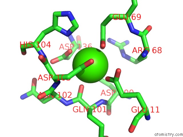
Mono view
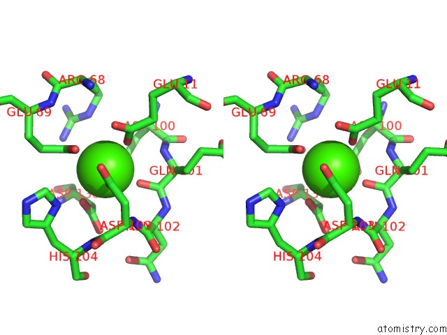
Stereo pair view
Mono view
Stereo pair view
A full contact list of Calcium with other atoms in the Ca binding
site number 1 of Crystal Structure of Human P-Cadherin (Ss-Dimer K14E) within 5.0Å range:
|
Calcium binding site 2 out of 2 in 4zmo
Go back to
Calcium binding site 2 out
of 2 in the Crystal Structure of Human P-Cadherin (Ss-Dimer K14E)
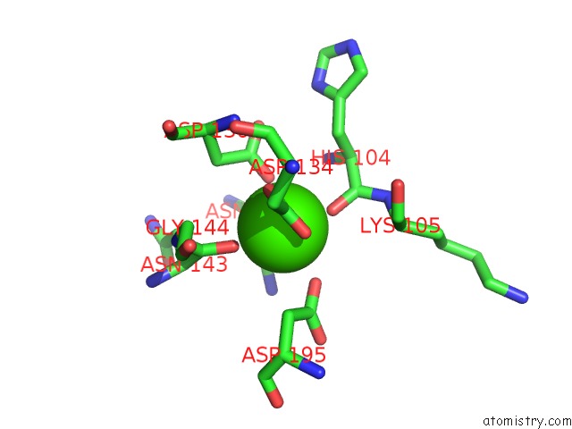
Mono view
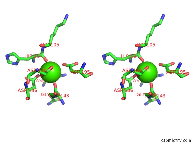
Stereo pair view
Mono view
Stereo pair view
A full contact list of Calcium with other atoms in the Ca binding
site number 2 of Crystal Structure of Human P-Cadherin (Ss-Dimer K14E) within 5.0Å range:
|
Reference:
S.Kudo,
J.M.Caaveiro,
K.Tsumoto.
Adhesive Dimerization of Human P-Cadherin Catalyzed By A Chaperone-Like Mechanism Structure V. 24 1523 2016.
ISSN: ISSN 0969-2126
PubMed: 27545624
DOI: 10.1016/J.STR.2016.07.002
Page generated: Wed Jul 9 03:45:05 2025
ISSN: ISSN 0969-2126
PubMed: 27545624
DOI: 10.1016/J.STR.2016.07.002
Last articles
Fe in 2YXOFe in 2YRS
Fe in 2YXC
Fe in 2YNM
Fe in 2YVJ
Fe in 2YP1
Fe in 2YU2
Fe in 2YU1
Fe in 2YQB
Fe in 2YOO