Calcium »
PDB 4zmq-5a3y »
4zvf »
Calcium in PDB 4zvf: Crystal Structure of Ggdef Domain of the E. Coli Dosc - Form II (Gtp- Alpha-S-Bound)
Enzymatic activity of Crystal Structure of Ggdef Domain of the E. Coli Dosc - Form II (Gtp- Alpha-S-Bound)
All present enzymatic activity of Crystal Structure of Ggdef Domain of the E. Coli Dosc - Form II (Gtp- Alpha-S-Bound):
2.7.7.65;
2.7.7.65;
Protein crystallography data
The structure of Crystal Structure of Ggdef Domain of the E. Coli Dosc - Form II (Gtp- Alpha-S-Bound), PDB code: 4zvf
was solved by
M.Tarnawski,
T.R.M.Barends,
I.Schlichting,
with X-Ray Crystallography technique. A brief refinement statistics is given in the table below:
| Resolution Low / High (Å) | 35.81 / 1.15 |
| Space group | P 1 21 1 |
| Cell size a, b, c (Å), α, β, γ (°) | 28.500, 52.530, 50.930, 90.00, 106.08, 90.00 |
| R / Rfree (%) | 13.8 / 16.5 |
Calcium Binding Sites:
The binding sites of Calcium atom in the Crystal Structure of Ggdef Domain of the E. Coli Dosc - Form II (Gtp- Alpha-S-Bound)
(pdb code 4zvf). This binding sites where shown within
5.0 Angstroms radius around Calcium atom.
In total 2 binding sites of Calcium where determined in the Crystal Structure of Ggdef Domain of the E. Coli Dosc - Form II (Gtp- Alpha-S-Bound), PDB code: 4zvf:
Jump to Calcium binding site number: 1; 2;
In total 2 binding sites of Calcium where determined in the Crystal Structure of Ggdef Domain of the E. Coli Dosc - Form II (Gtp- Alpha-S-Bound), PDB code: 4zvf:
Jump to Calcium binding site number: 1; 2;
Calcium binding site 1 out of 2 in 4zvf
Go back to
Calcium binding site 1 out
of 2 in the Crystal Structure of Ggdef Domain of the E. Coli Dosc - Form II (Gtp- Alpha-S-Bound)

Mono view

Stereo pair view
Mono view
Stereo pair view
A full contact list of Calcium with other atoms in the Ca binding
site number 1 of Crystal Structure of Ggdef Domain of the E. Coli Dosc - Form II (Gtp- Alpha-S-Bound) within 5.0Å range:
|
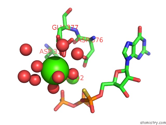
Calcium binding site 2 out of 2 in 4zvf
Go back to
Calcium binding site 2 out
of 2 in the Crystal Structure of Ggdef Domain of the E. Coli Dosc - Form II (Gtp- Alpha-S-Bound)
Mono view

Stereo pair view
Mono view
Stereo pair view
A full contact list of Calcium with other atoms in the Ca binding
site number 2 of Crystal Structure of Ggdef Domain of the E. Coli Dosc - Form II (Gtp- Alpha-S-Bound) within 5.0Å range:
|
Reference:
M.Tarnawski,
T.R.Barends,
I.Schlichting.
Structural Analysis of An Oxygen-Regulated Diguanylate Cyclase. Acta Crystallogr.,Sect.D V. 71 2158 2015.
ISSN: ESSN 1399-0047
PubMed: 26527135
DOI: 10.1107/S139900471501545X
Page generated: Wed Jul 9 03:58:13 2025
ISSN: ESSN 1399-0047
PubMed: 26527135
DOI: 10.1107/S139900471501545X
Last articles
Fe in 2YXOFe in 2YRS
Fe in 2YXC
Fe in 2YNM
Fe in 2YVJ
Fe in 2YP1
Fe in 2YU2
Fe in 2YU1
Fe in 2YQB
Fe in 2YOO