Calcium »
PDB 4zmq-5a3y »
5a2a »
Calcium in PDB 5a2a: Crystal Structure of Anoxybacillus Alpha-Amylase Provides Insights Into A New Glycosyl Hydrolase Subclass
Enzymatic activity of Crystal Structure of Anoxybacillus Alpha-Amylase Provides Insights Into A New Glycosyl Hydrolase Subclass
All present enzymatic activity of Crystal Structure of Anoxybacillus Alpha-Amylase Provides Insights Into A New Glycosyl Hydrolase Subclass:
3.2.1.1;
3.2.1.1;
Protein crystallography data
The structure of Crystal Structure of Anoxybacillus Alpha-Amylase Provides Insights Into A New Glycosyl Hydrolase Subclass, PDB code: 5a2a
was solved by
C.L.Ng,
K.P.Chai,
N.F.Othman,
A.H.Teh,
K.L.Ho,
K.G.Chan,
K.M.Goh,
with X-Ray Crystallography technique. A brief refinement statistics is given in the table below:
| Resolution Low / High (Å) | 19.99 / 1.90 |
| Space group | P 21 21 21 |
| Cell size a, b, c (Å), α, β, γ (°) | 61.429, 63.443, 122.758, 90.00, 90.00, 90.00 |
| R / Rfree (%) | 17.689 / 23.6 |
Calcium Binding Sites:
The binding sites of Calcium atom in the Crystal Structure of Anoxybacillus Alpha-Amylase Provides Insights Into A New Glycosyl Hydrolase Subclass
(pdb code 5a2a). This binding sites where shown within
5.0 Angstroms radius around Calcium atom.
In total 4 binding sites of Calcium where determined in the Crystal Structure of Anoxybacillus Alpha-Amylase Provides Insights Into A New Glycosyl Hydrolase Subclass, PDB code: 5a2a:
Jump to Calcium binding site number: 1; 2; 3; 4;
In total 4 binding sites of Calcium where determined in the Crystal Structure of Anoxybacillus Alpha-Amylase Provides Insights Into A New Glycosyl Hydrolase Subclass, PDB code: 5a2a:
Jump to Calcium binding site number: 1; 2; 3; 4;
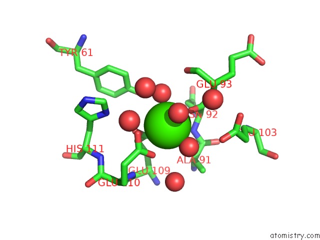
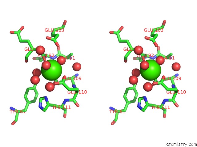
Calcium binding site 1 out of 4 in 5a2a
Go back to
Calcium binding site 1 out
of 4 in the Crystal Structure of Anoxybacillus Alpha-Amylase Provides Insights Into A New Glycosyl Hydrolase Subclass
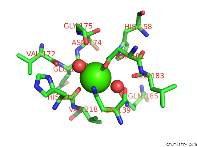
Mono view
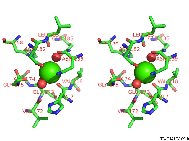
Stereo pair view
Mono view
Stereo pair view
A full contact list of Calcium with other atoms in the Ca binding
site number 1 of Crystal Structure of Anoxybacillus Alpha-Amylase Provides Insights Into A New Glycosyl Hydrolase Subclass within 5.0Å range:
|
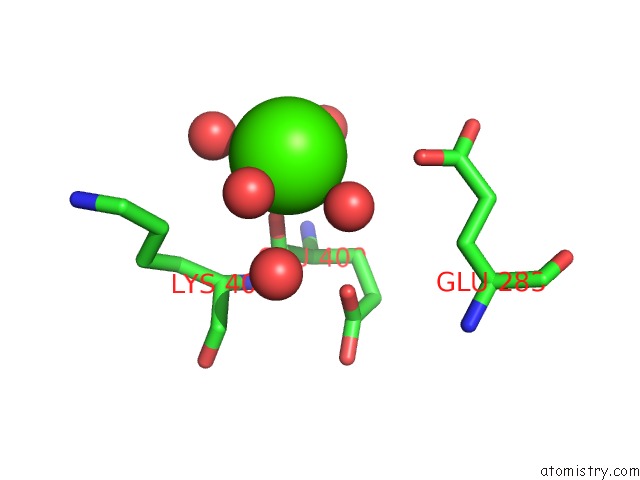
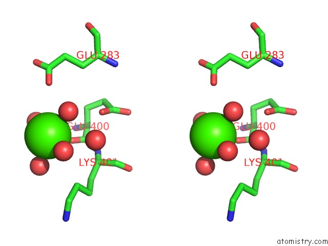
Calcium binding site 2 out of 4 in 5a2a
Go back to
Calcium binding site 2 out
of 4 in the Crystal Structure of Anoxybacillus Alpha-Amylase Provides Insights Into A New Glycosyl Hydrolase Subclass
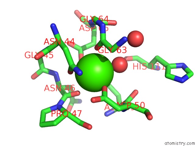
Mono view
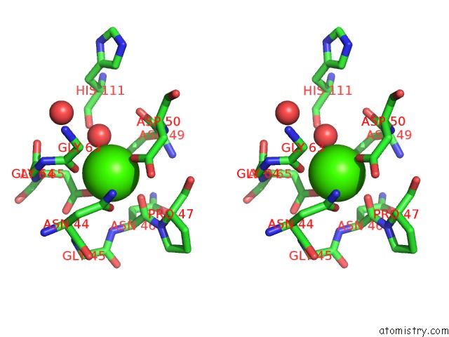
Stereo pair view
Mono view
Stereo pair view
A full contact list of Calcium with other atoms in the Ca binding
site number 2 of Crystal Structure of Anoxybacillus Alpha-Amylase Provides Insights Into A New Glycosyl Hydrolase Subclass within 5.0Å range:
|
Calcium binding site 3 out of 4 in 5a2a
Go back to
Calcium binding site 3 out
of 4 in the Crystal Structure of Anoxybacillus Alpha-Amylase Provides Insights Into A New Glycosyl Hydrolase Subclass
Mono view
Stereo pair view
Mono view
Stereo pair view
A full contact list of Calcium with other atoms in the Ca binding
site number 3 of Crystal Structure of Anoxybacillus Alpha-Amylase Provides Insights Into A New Glycosyl Hydrolase Subclass within 5.0Å range:
|
Calcium binding site 4 out of 4 in 5a2a
Go back to
Calcium binding site 4 out
of 4 in the Crystal Structure of Anoxybacillus Alpha-Amylase Provides Insights Into A New Glycosyl Hydrolase Subclass
Mono view
Stereo pair view
Mono view
Stereo pair view
A full contact list of Calcium with other atoms in the Ca binding
site number 4 of Crystal Structure of Anoxybacillus Alpha-Amylase Provides Insights Into A New Glycosyl Hydrolase Subclass within 5.0Å range:
|
Reference:
K.P.Chai,
N.F.B.Othman,
A.Teh,
K.L.Ho,
K.Chan,
M.S.Shamsir,
K.M.Goh,
C.L.Ng.
Crystal Structure of Anoxybacillus Alpha-Amylase Provides Insights Into Maltose Binding of A New Glycosyl Hydrolase Subclass. Sci.Rep. V. 6 23126 2016.
ISSN: ISSN 2045-2322
PubMed: 26975884
DOI: 10.1038/SREP23126
Page generated: Wed Jul 9 03:59:03 2025
ISSN: ISSN 2045-2322
PubMed: 26975884
DOI: 10.1038/SREP23126
Last articles
Fe in 2YXOFe in 2YRS
Fe in 2YXC
Fe in 2YNM
Fe in 2YVJ
Fe in 2YP1
Fe in 2YU2
Fe in 2YU1
Fe in 2YQB
Fe in 2YOO