Calcium »
PDB 5a4x-5afp »
5a56 »
Calcium in PDB 5a56: The Structure of GH101 From Streptococcus Pneumoniae TIGR4 in Complex with 1-O-Methyl-T-Antigen
Enzymatic activity of The Structure of GH101 From Streptococcus Pneumoniae TIGR4 in Complex with 1-O-Methyl-T-Antigen
All present enzymatic activity of The Structure of GH101 From Streptococcus Pneumoniae TIGR4 in Complex with 1-O-Methyl-T-Antigen:
3.2.1.97;
3.2.1.97;
Protein crystallography data
The structure of The Structure of GH101 From Streptococcus Pneumoniae TIGR4 in Complex with 1-O-Methyl-T-Antigen, PDB code: 5a56
was solved by
K.J.Gregg,
M.D.L.Suits,
L.Deng,
D.J.Vocadlo,
A.B.Boraston,
with X-Ray Crystallography technique. A brief refinement statistics is given in the table below:
| Resolution Low / High (Å) | 43.42 / 1.80 |
| Space group | P 2 21 21 |
| Cell size a, b, c (Å), α, β, γ (°) | 86.713, 121.764, 139.429, 90.00, 90.00, 90.00 |
| R / Rfree (%) | 15.2 / 17.8 |
Other elements in 5a56:
The structure of The Structure of GH101 From Streptococcus Pneumoniae TIGR4 in Complex with 1-O-Methyl-T-Antigen also contains other interesting chemical elements:
| Manganese | (Mn) | 1 atom |
Calcium Binding Sites:
The binding sites of Calcium atom in the The Structure of GH101 From Streptococcus Pneumoniae TIGR4 in Complex with 1-O-Methyl-T-Antigen
(pdb code 5a56). This binding sites where shown within
5.0 Angstroms radius around Calcium atom.
In total 3 binding sites of Calcium where determined in the The Structure of GH101 From Streptococcus Pneumoniae TIGR4 in Complex with 1-O-Methyl-T-Antigen, PDB code: 5a56:
Jump to Calcium binding site number: 1; 2; 3;
In total 3 binding sites of Calcium where determined in the The Structure of GH101 From Streptococcus Pneumoniae TIGR4 in Complex with 1-O-Methyl-T-Antigen, PDB code: 5a56:
Jump to Calcium binding site number: 1; 2; 3;
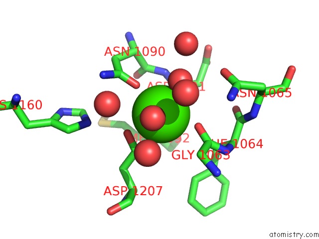
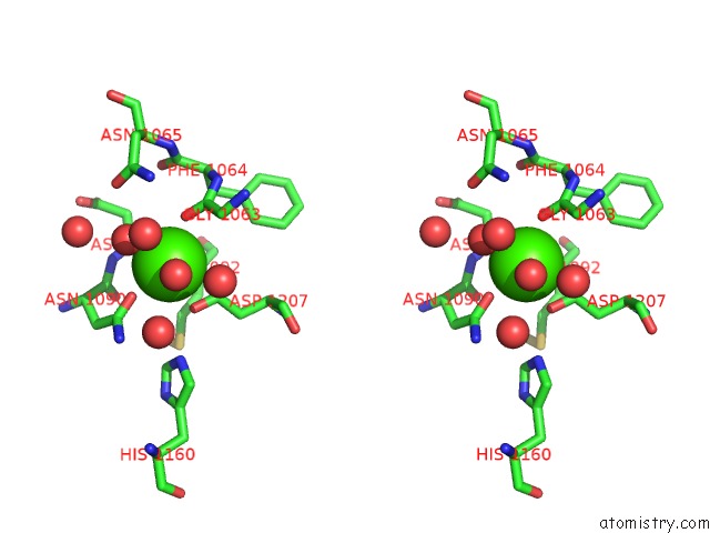
Calcium binding site 1 out of 3 in 5a56
Go back to
Calcium binding site 1 out
of 3 in the The Structure of GH101 From Streptococcus Pneumoniae TIGR4 in Complex with 1-O-Methyl-T-Antigen
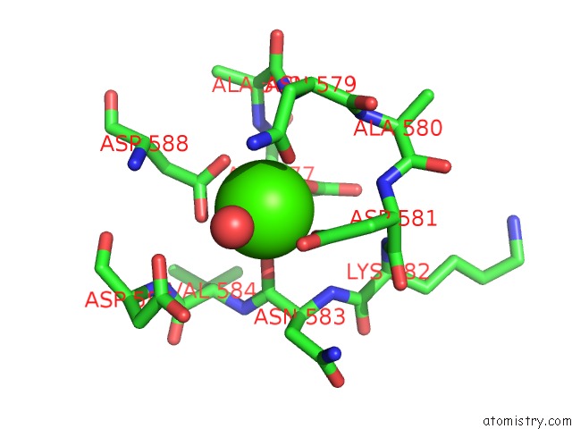
Mono view
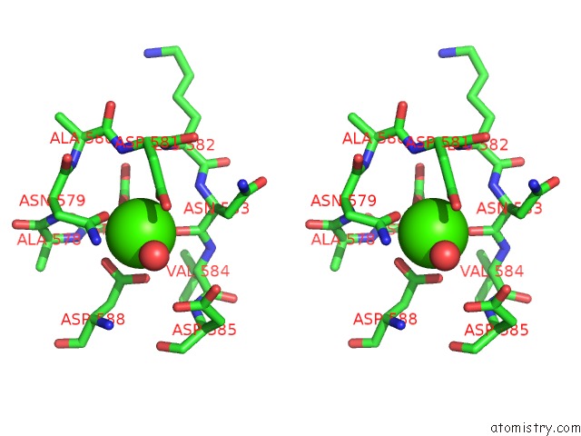
Stereo pair view
Mono view
Stereo pair view
A full contact list of Calcium with other atoms in the Ca binding
site number 1 of The Structure of GH101 From Streptococcus Pneumoniae TIGR4 in Complex with 1-O-Methyl-T-Antigen within 5.0Å range:
|
Calcium binding site 2 out of 3 in 5a56
Go back to
Calcium binding site 2 out
of 3 in the The Structure of GH101 From Streptococcus Pneumoniae TIGR4 in Complex with 1-O-Methyl-T-Antigen
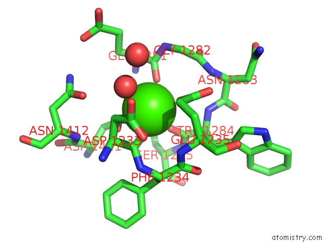
Mono view
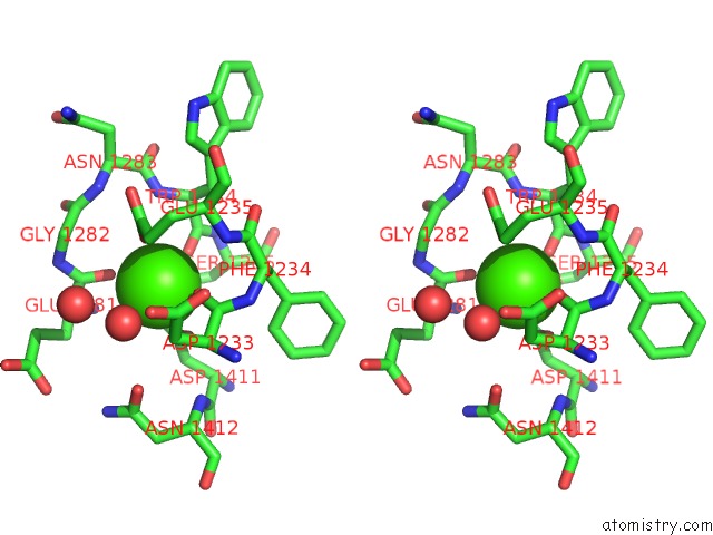
Stereo pair view
Mono view
Stereo pair view
A full contact list of Calcium with other atoms in the Ca binding
site number 2 of The Structure of GH101 From Streptococcus Pneumoniae TIGR4 in Complex with 1-O-Methyl-T-Antigen within 5.0Å range:
|
Calcium binding site 3 out of 3 in 5a56
Go back to
Calcium binding site 3 out
of 3 in the The Structure of GH101 From Streptococcus Pneumoniae TIGR4 in Complex with 1-O-Methyl-T-Antigen
Mono view
Stereo pair view
Mono view
Stereo pair view
A full contact list of Calcium with other atoms in the Ca binding
site number 3 of The Structure of GH101 From Streptococcus Pneumoniae TIGR4 in Complex with 1-O-Methyl-T-Antigen within 5.0Å range:
|
Reference:
K.J.Gregg,
M.D.Suits,
L.Deng,
D.J.Vocadlo,
A.B.Boraston.
Structural Analysis of A Family 101 Glycoside Hydrolase in Complex with Carbohydrates Reveals Insights Into Its Mechanism. J.Biol.Chem. V. 290 25657 2015.
ISSN: ESSN 1083-351X
PubMed: 26304114
DOI: 10.1074/JBC.M115.680470
Page generated: Wed Jul 9 04:03:12 2025
ISSN: ESSN 1083-351X
PubMed: 26304114
DOI: 10.1074/JBC.M115.680470
Last articles
Fe in 2YXOFe in 2YRS
Fe in 2YXC
Fe in 2YNM
Fe in 2YVJ
Fe in 2YP1
Fe in 2YU2
Fe in 2YU1
Fe in 2YQB
Fe in 2YOO