Calcium »
PDB 5a4x-5afp »
5a6x »
Calcium in PDB 5a6x: Structure of the Lecb Lectin From Pseudomonas Aeruginosa Strain PA14 in Complex with Alpha-Methyl-Fucoside
Protein crystallography data
The structure of Structure of the Lecb Lectin From Pseudomonas Aeruginosa Strain PA14 in Complex with Alpha-Methyl-Fucoside, PDB code: 5a6x
was solved by
R.Sommer,
S.Wagner,
A.Varrot,
A.Khaledi,
S.Haussler,
A.Imberty,
A.Titz,
with X-Ray Crystallography technique. A brief refinement statistics is given in the table below:
| Resolution Low / High (Å) | 37.84 / 1.55 |
| Space group | P 21 21 21 |
| Cell size a, b, c (Å), α, β, γ (°) | 52.558, 65.585, 109.036, 90.00, 90.00, 90.00 |
| R / Rfree (%) | 12.8 / 15.3 |
Calcium Binding Sites:
The binding sites of Calcium atom in the Structure of the Lecb Lectin From Pseudomonas Aeruginosa Strain PA14 in Complex with Alpha-Methyl-Fucoside
(pdb code 5a6x). This binding sites where shown within
5.0 Angstroms radius around Calcium atom.
In total 8 binding sites of Calcium where determined in the Structure of the Lecb Lectin From Pseudomonas Aeruginosa Strain PA14 in Complex with Alpha-Methyl-Fucoside, PDB code: 5a6x:
Jump to Calcium binding site number: 1; 2; 3; 4; 5; 6; 7; 8;
In total 8 binding sites of Calcium where determined in the Structure of the Lecb Lectin From Pseudomonas Aeruginosa Strain PA14 in Complex with Alpha-Methyl-Fucoside, PDB code: 5a6x:
Jump to Calcium binding site number: 1; 2; 3; 4; 5; 6; 7; 8;
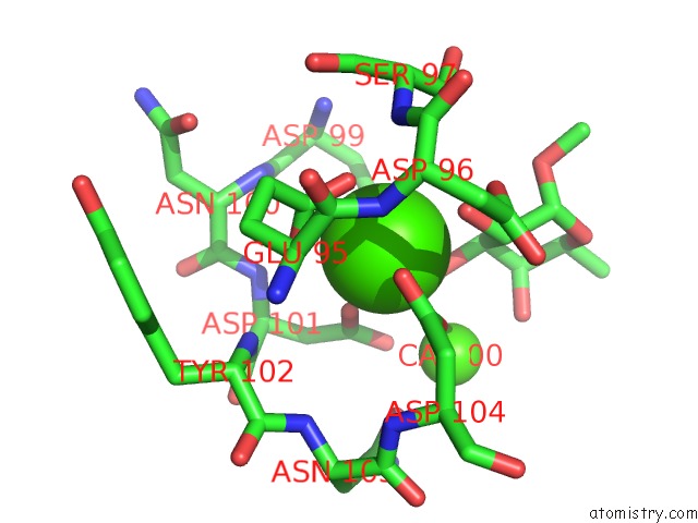
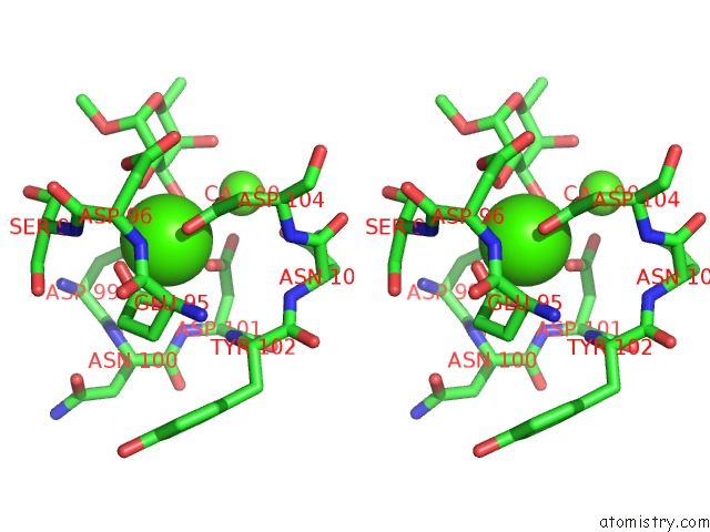
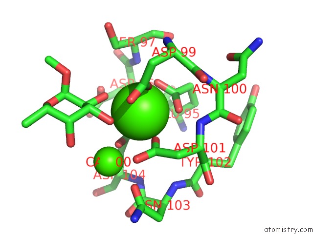
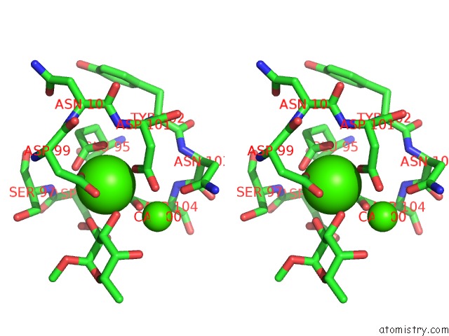
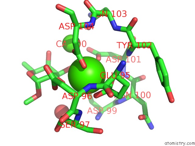
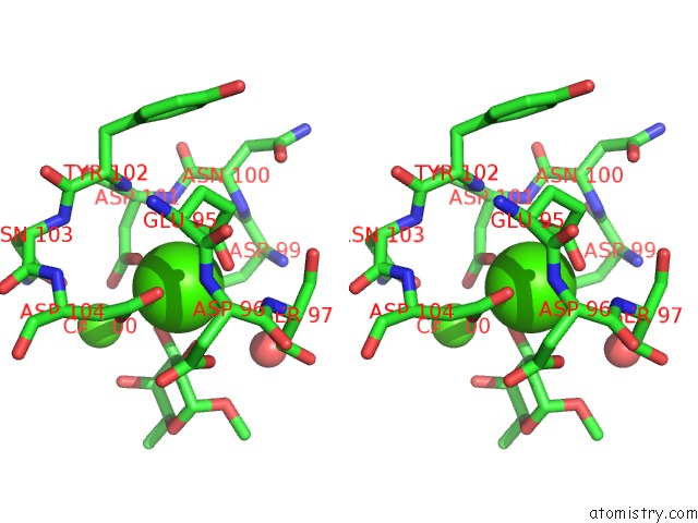
Calcium binding site 1 out of 8 in 5a6x
Go back to
Calcium binding site 1 out
of 8 in the Structure of the Lecb Lectin From Pseudomonas Aeruginosa Strain PA14 in Complex with Alpha-Methyl-Fucoside
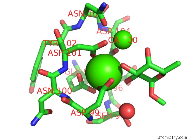
Mono view
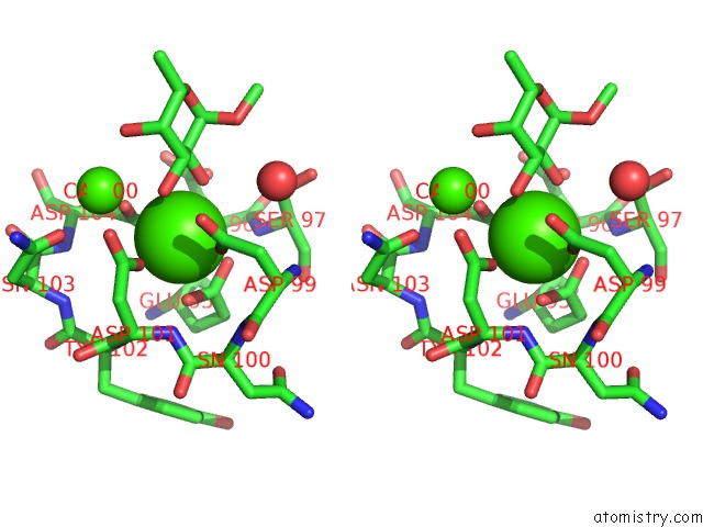
Stereo pair view
Mono view
Stereo pair view
A full contact list of Calcium with other atoms in the Ca binding
site number 1 of Structure of the Lecb Lectin From Pseudomonas Aeruginosa Strain PA14 in Complex with Alpha-Methyl-Fucoside within 5.0Å range:
|
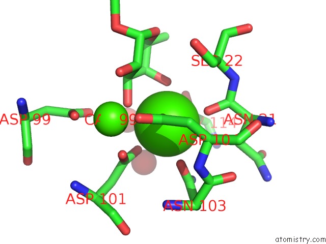
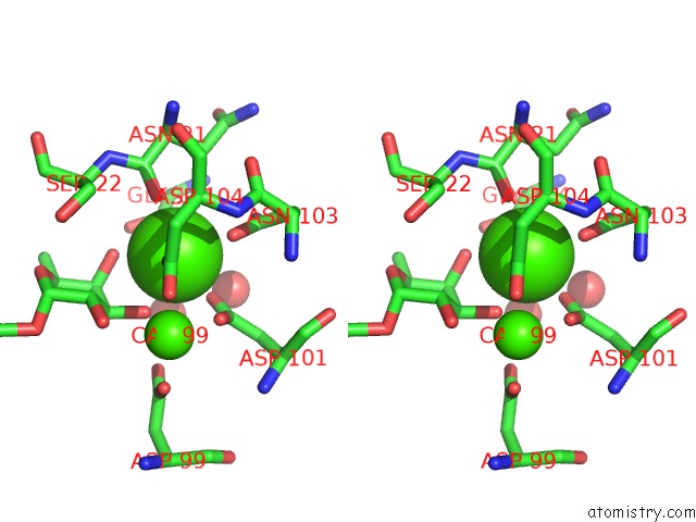
Calcium binding site 2 out of 8 in 5a6x
Go back to
Calcium binding site 2 out
of 8 in the Structure of the Lecb Lectin From Pseudomonas Aeruginosa Strain PA14 in Complex with Alpha-Methyl-Fucoside
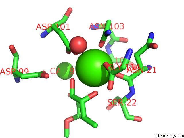
Mono view
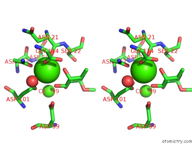
Stereo pair view
Mono view
Stereo pair view
A full contact list of Calcium with other atoms in the Ca binding
site number 2 of Structure of the Lecb Lectin From Pseudomonas Aeruginosa Strain PA14 in Complex with Alpha-Methyl-Fucoside within 5.0Å range:
|
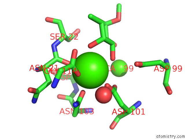
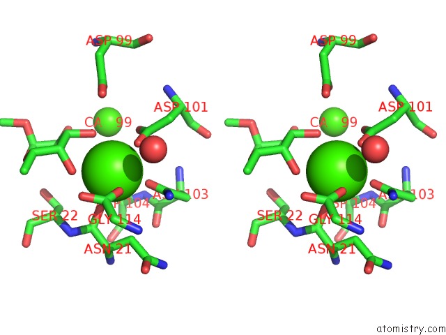
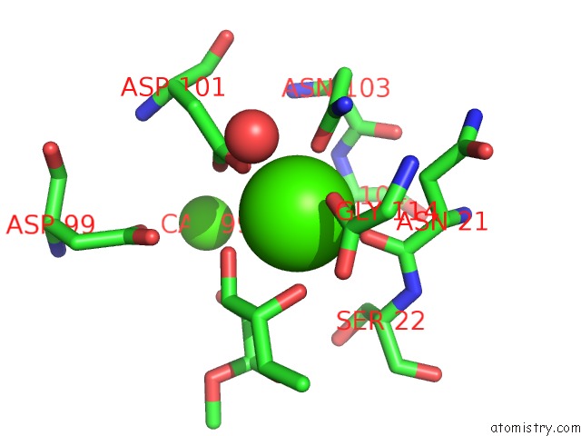
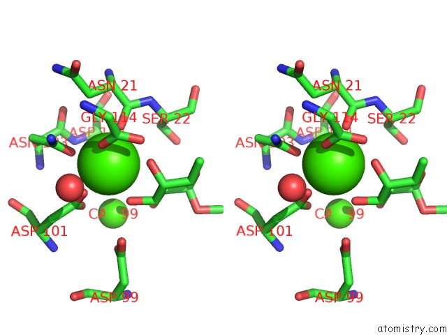
Calcium binding site 3 out of 8 in 5a6x
Go back to
Calcium binding site 3 out
of 8 in the Structure of the Lecb Lectin From Pseudomonas Aeruginosa Strain PA14 in Complex with Alpha-Methyl-Fucoside
Mono view
Stereo pair view
Mono view
Stereo pair view
A full contact list of Calcium with other atoms in the Ca binding
site number 3 of Structure of the Lecb Lectin From Pseudomonas Aeruginosa Strain PA14 in Complex with Alpha-Methyl-Fucoside within 5.0Å range:
|
Calcium binding site 4 out of 8 in 5a6x
Go back to
Calcium binding site 4 out
of 8 in the Structure of the Lecb Lectin From Pseudomonas Aeruginosa Strain PA14 in Complex with Alpha-Methyl-Fucoside
Mono view
Stereo pair view
Mono view
Stereo pair view
A full contact list of Calcium with other atoms in the Ca binding
site number 4 of Structure of the Lecb Lectin From Pseudomonas Aeruginosa Strain PA14 in Complex with Alpha-Methyl-Fucoside within 5.0Å range:
|
Calcium binding site 5 out of 8 in 5a6x
Go back to
Calcium binding site 5 out
of 8 in the Structure of the Lecb Lectin From Pseudomonas Aeruginosa Strain PA14 in Complex with Alpha-Methyl-Fucoside
Mono view
Stereo pair view
Mono view
Stereo pair view
A full contact list of Calcium with other atoms in the Ca binding
site number 5 of Structure of the Lecb Lectin From Pseudomonas Aeruginosa Strain PA14 in Complex with Alpha-Methyl-Fucoside within 5.0Å range:
|
Calcium binding site 6 out of 8 in 5a6x
Go back to
Calcium binding site 6 out
of 8 in the Structure of the Lecb Lectin From Pseudomonas Aeruginosa Strain PA14 in Complex with Alpha-Methyl-Fucoside
Mono view
Stereo pair view
Mono view
Stereo pair view
A full contact list of Calcium with other atoms in the Ca binding
site number 6 of Structure of the Lecb Lectin From Pseudomonas Aeruginosa Strain PA14 in Complex with Alpha-Methyl-Fucoside within 5.0Å range:
|
Calcium binding site 7 out of 8 in 5a6x
Go back to
Calcium binding site 7 out
of 8 in the Structure of the Lecb Lectin From Pseudomonas Aeruginosa Strain PA14 in Complex with Alpha-Methyl-Fucoside
Mono view
Stereo pair view
Mono view
Stereo pair view
A full contact list of Calcium with other atoms in the Ca binding
site number 7 of Structure of the Lecb Lectin From Pseudomonas Aeruginosa Strain PA14 in Complex with Alpha-Methyl-Fucoside within 5.0Å range:
|
Calcium binding site 8 out of 8 in 5a6x
Go back to
Calcium binding site 8 out
of 8 in the Structure of the Lecb Lectin From Pseudomonas Aeruginosa Strain PA14 in Complex with Alpha-Methyl-Fucoside
Mono view
Stereo pair view
Mono view
Stereo pair view
A full contact list of Calcium with other atoms in the Ca binding
site number 8 of Structure of the Lecb Lectin From Pseudomonas Aeruginosa Strain PA14 in Complex with Alpha-Methyl-Fucoside within 5.0Å range:
|
Reference:
R.Sommer,
S.Wagner,
A.Varrot,
C.M.Nycholat,
A.Khaledi,
S.Haussler,
J.C.Paulson,
A.Imberty,
A.Titz.
The Virulence Factor Lecb Varies in Clinical Isolates: Consequences For Ligand Binding and Drug Discovery. Chem Sci V. 7 4990 2016.
ISSN: ISSN 2041-6520
PubMed: 30155149
DOI: 10.1039/C6SC00696E
Page generated: Wed Jul 9 04:04:14 2025
ISSN: ISSN 2041-6520
PubMed: 30155149
DOI: 10.1039/C6SC00696E
Last articles
F in 7LCOF in 7LCK
F in 7LCJ
F in 7LCI
F in 7L9Y
F in 7LCD
F in 7LAY
F in 7LAJ
F in 7LAE
F in 7L9M