Calcium »
PDB 5a4x-5afp »
5afb »
Calcium in PDB 5afb: Crystal Structure of the LATROPHILIN3 Lectin and Olfactomedin Domains
Protein crystallography data
The structure of Crystal Structure of the LATROPHILIN3 Lectin and Olfactomedin Domains, PDB code: 5afb
was solved by
V.A.Jackson,
D.Del Toro,
M.Carrasquero,
P.Roversi,
K.Harlos,
R.Klein,
E.Seiradake,
with X-Ray Crystallography technique. A brief refinement statistics is given in the table below:
| Resolution Low / High (Å) | 22.91 / 2.16 |
| Space group | I 2 2 2 |
| Cell size a, b, c (Å), α, β, γ (°) | 78.430, 96.650, 101.640, 90.00, 90.00, 90.00 |
| R / Rfree (%) | 20.44 / 24.52 |
Other elements in 5afb:
The structure of Crystal Structure of the LATROPHILIN3 Lectin and Olfactomedin Domains also contains other interesting chemical elements:
| Sodium | (Na) | 1 atom |
Calcium Binding Sites:
The binding sites of Calcium atom in the Crystal Structure of the LATROPHILIN3 Lectin and Olfactomedin Domains
(pdb code 5afb). This binding sites where shown within
5.0 Angstroms radius around Calcium atom.
In total only one binding site of Calcium was determined in the Crystal Structure of the LATROPHILIN3 Lectin and Olfactomedin Domains, PDB code: 5afb:
In total only one binding site of Calcium was determined in the Crystal Structure of the LATROPHILIN3 Lectin and Olfactomedin Domains, PDB code: 5afb:
Calcium binding site 1 out of 1 in 5afb
Go back to
Calcium binding site 1 out
of 1 in the Crystal Structure of the LATROPHILIN3 Lectin and Olfactomedin Domains
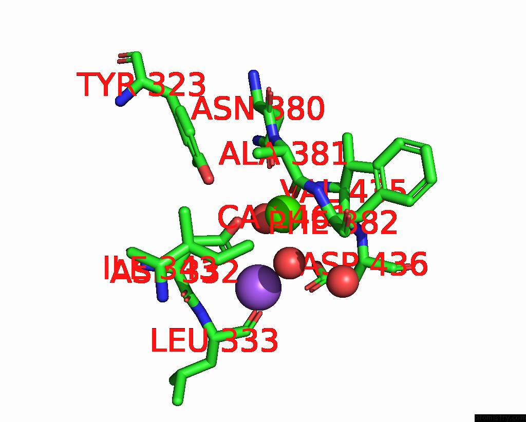
Mono view
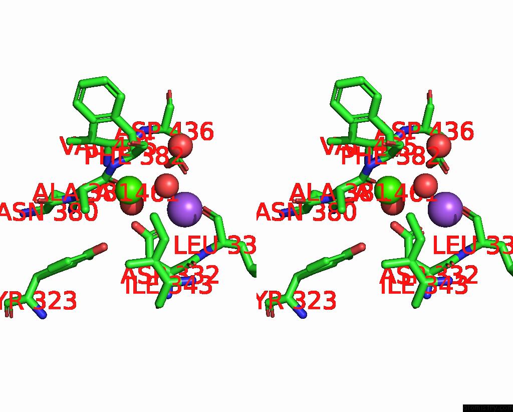
Stereo pair view
Mono view
Stereo pair view
A full contact list of Calcium with other atoms in the Ca binding
site number 1 of Crystal Structure of the LATROPHILIN3 Lectin and Olfactomedin Domains within 5.0Å range:
|
Reference:
V.A.Jackson,
D.Del Toro,
M.Carrasquero,
P.Roversi,
K.Harlos,
R.Klein,
E.Seiradake.
Structural Basis of Latrophilin-Flrt Interaction. Structure 2015.
ISSN: ESSN 1878-4186
PubMed: 25728924
DOI: 10.1016/J.STR.2015.01.013
Page generated: Wed Jul 9 04:11:30 2025
ISSN: ESSN 1878-4186
PubMed: 25728924
DOI: 10.1016/J.STR.2015.01.013
Last articles
Fe in 2YXOFe in 2YRS
Fe in 2YXC
Fe in 2YNM
Fe in 2YVJ
Fe in 2YP1
Fe in 2YU2
Fe in 2YU1
Fe in 2YQB
Fe in 2YOO