Calcium »
PDB 5agv-5b4y »
5aza »
Calcium in PDB 5aza: Crystal Structure of Mbp-Saglb Fusion Protein with A 20-Residue Spacer in the Connector Helix
Protein crystallography data
The structure of Crystal Structure of Mbp-Saglb Fusion Protein with A 20-Residue Spacer in the Connector Helix, PDB code: 5aza
was solved by
R.Matsuoka,
D.Kohda,
with X-Ray Crystallography technique. A brief refinement statistics is given in the table below:
| Resolution Low / High (Å) | 50.00 / 2.08 |
| Space group | P 21 21 21 |
| Cell size a, b, c (Å), α, β, γ (°) | 66.468, 100.467, 140.920, 90.00, 90.00, 90.00 |
| R / Rfree (%) | 19.6 / 24.8 |
Calcium Binding Sites:
The binding sites of Calcium atom in the Crystal Structure of Mbp-Saglb Fusion Protein with A 20-Residue Spacer in the Connector Helix
(pdb code 5aza). This binding sites where shown within
5.0 Angstroms radius around Calcium atom.
In total only one binding site of Calcium was determined in the Crystal Structure of Mbp-Saglb Fusion Protein with A 20-Residue Spacer in the Connector Helix, PDB code: 5aza:
In total only one binding site of Calcium was determined in the Crystal Structure of Mbp-Saglb Fusion Protein with A 20-Residue Spacer in the Connector Helix, PDB code: 5aza:
Calcium binding site 1 out of 1 in 5aza
Go back to
Calcium binding site 1 out
of 1 in the Crystal Structure of Mbp-Saglb Fusion Protein with A 20-Residue Spacer in the Connector Helix
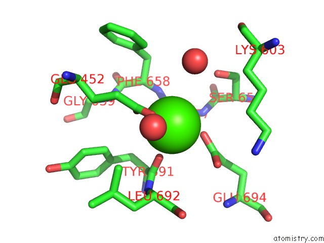
Mono view
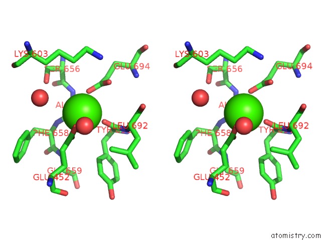
Stereo pair view
Mono view
Stereo pair view
A full contact list of Calcium with other atoms in the Ca binding
site number 1 of Crystal Structure of Mbp-Saglb Fusion Protein with A 20-Residue Spacer in the Connector Helix within 5.0Å range:
|
Reference:
R.Matsuoka,
A.Shimada,
Y.Komuro,
Y.Sugita,
D.Kohda.
Rational Design of Crystal Contact-Free Space in Protein Crystals For Analyzing Spatial Distribution of Motions Within Protein Molecules. Protein Sci. V. 25 754 2016.
ISSN: ESSN 1469-896X
PubMed: 26694222
DOI: 10.1002/PRO.2867
Page generated: Sun Jul 14 16:39:13 2024
ISSN: ESSN 1469-896X
PubMed: 26694222
DOI: 10.1002/PRO.2867
Last articles
Zn in 9J0NZn in 9J0O
Zn in 9J0P
Zn in 9FJX
Zn in 9EKB
Zn in 9C0F
Zn in 9CAH
Zn in 9CH0
Zn in 9CH3
Zn in 9CH1