Calcium »
PDB 5agv-5b4y »
5b1w »
Calcium in PDB 5b1w: Crystal Structure of Human Dendritic Cell Inhibitory Receptor (Dcir) C-Type Lectin Domain in Ligand-Free Form
Protein crystallography data
The structure of Crystal Structure of Human Dendritic Cell Inhibitory Receptor (Dcir) C-Type Lectin Domain in Ligand-Free Form, PDB code: 5b1w
was solved by
M.Nagae,
Y.Yamaguchi,
with X-Ray Crystallography technique. A brief refinement statistics is given in the table below:
| Resolution Low / High (Å) | 47.02 / 3.05 |
| Space group | P 21 21 2 |
| Cell size a, b, c (Å), α, β, γ (°) | 102.489, 105.832, 65.387, 90.00, 90.00, 90.00 |
| R / Rfree (%) | 25.3 / 28.5 |
Calcium Binding Sites:
The binding sites of Calcium atom in the Crystal Structure of Human Dendritic Cell Inhibitory Receptor (Dcir) C-Type Lectin Domain in Ligand-Free Form
(pdb code 5b1w). This binding sites where shown within
5.0 Angstroms radius around Calcium atom.
In total 8 binding sites of Calcium where determined in the Crystal Structure of Human Dendritic Cell Inhibitory Receptor (Dcir) C-Type Lectin Domain in Ligand-Free Form, PDB code: 5b1w:
Jump to Calcium binding site number: 1; 2; 3; 4; 5; 6; 7; 8;
In total 8 binding sites of Calcium where determined in the Crystal Structure of Human Dendritic Cell Inhibitory Receptor (Dcir) C-Type Lectin Domain in Ligand-Free Form, PDB code: 5b1w:
Jump to Calcium binding site number: 1; 2; 3; 4; 5; 6; 7; 8;
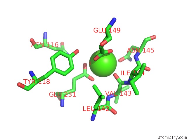
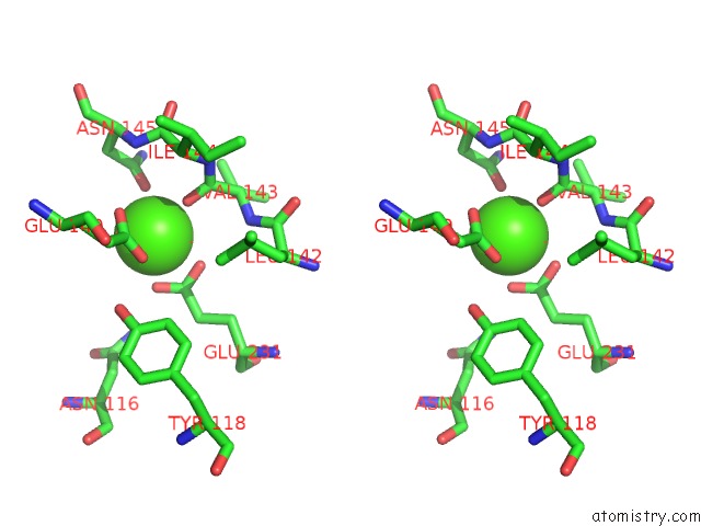
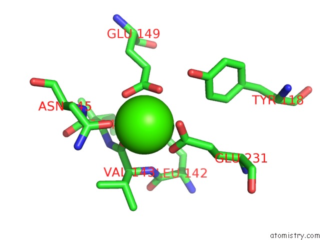
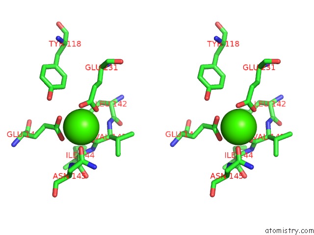
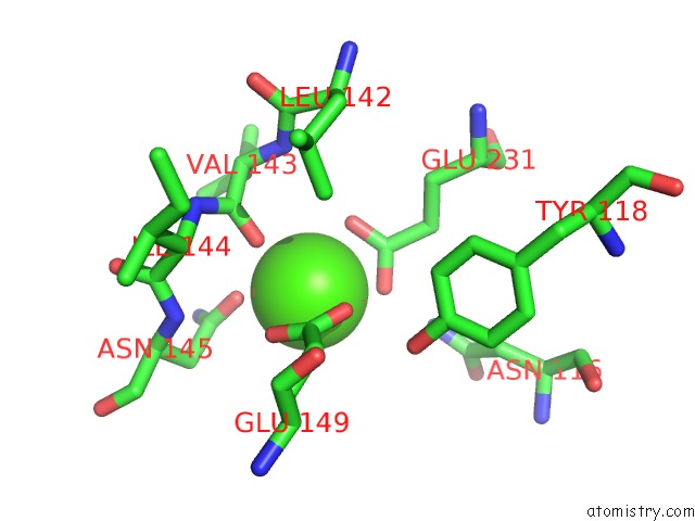
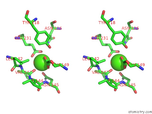
Calcium binding site 1 out of 8 in 5b1w
Go back to
Calcium binding site 1 out
of 8 in the Crystal Structure of Human Dendritic Cell Inhibitory Receptor (Dcir) C-Type Lectin Domain in Ligand-Free Form
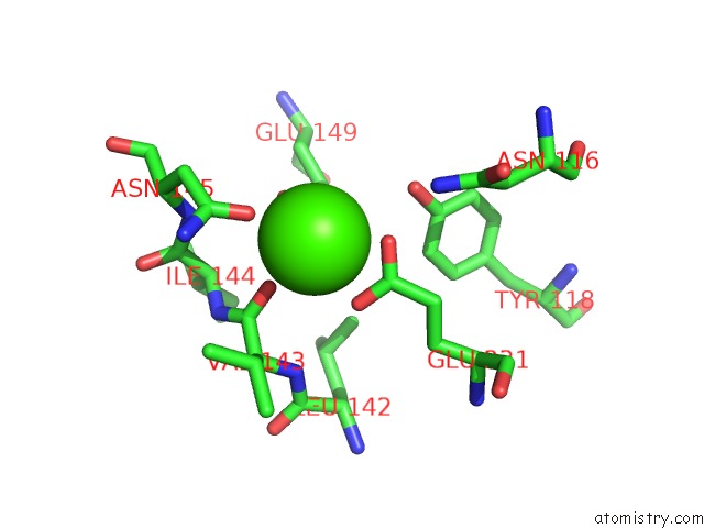
Mono view
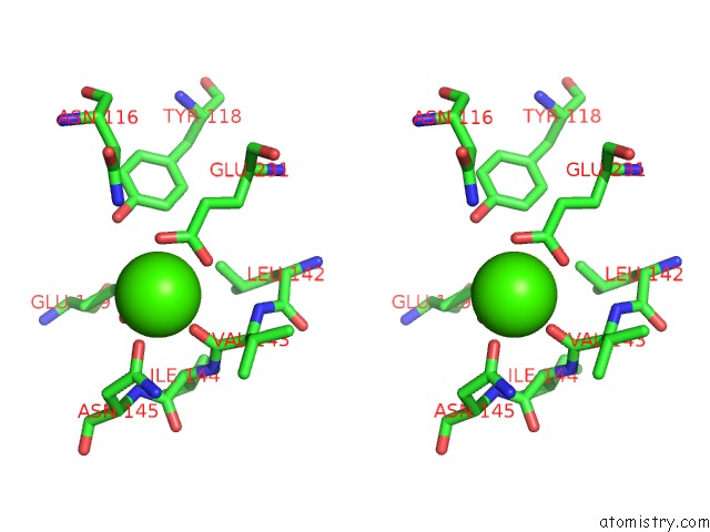
Stereo pair view
Mono view
Stereo pair view
A full contact list of Calcium with other atoms in the Ca binding
site number 1 of Crystal Structure of Human Dendritic Cell Inhibitory Receptor (Dcir) C-Type Lectin Domain in Ligand-Free Form within 5.0Å range:
|
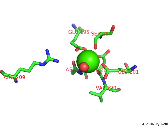
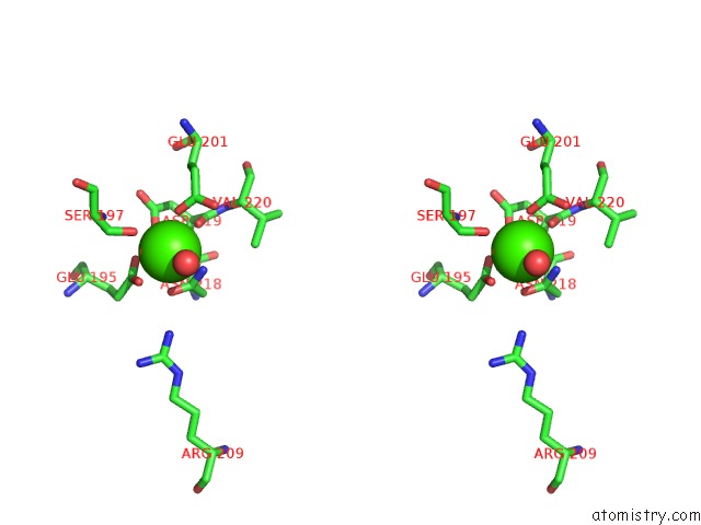
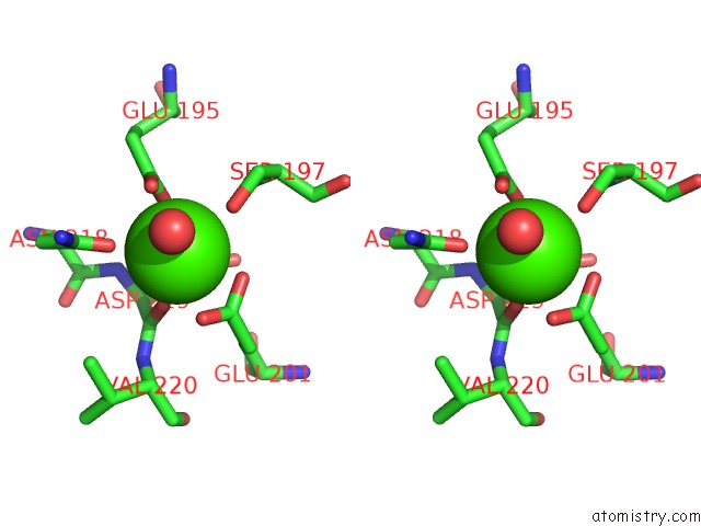
Calcium binding site 2 out of 8 in 5b1w
Go back to
Calcium binding site 2 out
of 8 in the Crystal Structure of Human Dendritic Cell Inhibitory Receptor (Dcir) C-Type Lectin Domain in Ligand-Free Form
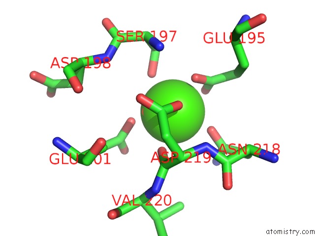
Mono view
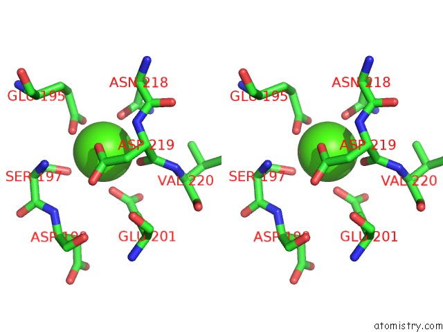
Stereo pair view
Mono view
Stereo pair view
A full contact list of Calcium with other atoms in the Ca binding
site number 2 of Crystal Structure of Human Dendritic Cell Inhibitory Receptor (Dcir) C-Type Lectin Domain in Ligand-Free Form within 5.0Å range:
|
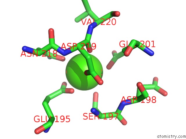
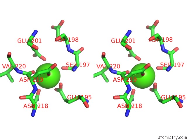
Calcium binding site 3 out of 8 in 5b1w
Go back to
Calcium binding site 3 out
of 8 in the Crystal Structure of Human Dendritic Cell Inhibitory Receptor (Dcir) C-Type Lectin Domain in Ligand-Free Form
Mono view
Stereo pair view
Mono view
Stereo pair view
A full contact list of Calcium with other atoms in the Ca binding
site number 3 of Crystal Structure of Human Dendritic Cell Inhibitory Receptor (Dcir) C-Type Lectin Domain in Ligand-Free Form within 5.0Å range:
|
Calcium binding site 4 out of 8 in 5b1w
Go back to
Calcium binding site 4 out
of 8 in the Crystal Structure of Human Dendritic Cell Inhibitory Receptor (Dcir) C-Type Lectin Domain in Ligand-Free Form
Mono view
Stereo pair view
Mono view
Stereo pair view
A full contact list of Calcium with other atoms in the Ca binding
site number 4 of Crystal Structure of Human Dendritic Cell Inhibitory Receptor (Dcir) C-Type Lectin Domain in Ligand-Free Form within 5.0Å range:
|
Calcium binding site 5 out of 8 in 5b1w
Go back to
Calcium binding site 5 out
of 8 in the Crystal Structure of Human Dendritic Cell Inhibitory Receptor (Dcir) C-Type Lectin Domain in Ligand-Free Form
Mono view
Stereo pair view
Mono view
Stereo pair view
A full contact list of Calcium with other atoms in the Ca binding
site number 5 of Crystal Structure of Human Dendritic Cell Inhibitory Receptor (Dcir) C-Type Lectin Domain in Ligand-Free Form within 5.0Å range:
|
Calcium binding site 6 out of 8 in 5b1w
Go back to
Calcium binding site 6 out
of 8 in the Crystal Structure of Human Dendritic Cell Inhibitory Receptor (Dcir) C-Type Lectin Domain in Ligand-Free Form
Mono view
Stereo pair view
Mono view
Stereo pair view
A full contact list of Calcium with other atoms in the Ca binding
site number 6 of Crystal Structure of Human Dendritic Cell Inhibitory Receptor (Dcir) C-Type Lectin Domain in Ligand-Free Form within 5.0Å range:
|
Calcium binding site 7 out of 8 in 5b1w
Go back to
Calcium binding site 7 out
of 8 in the Crystal Structure of Human Dendritic Cell Inhibitory Receptor (Dcir) C-Type Lectin Domain in Ligand-Free Form
Mono view
Stereo pair view
Mono view
Stereo pair view
A full contact list of Calcium with other atoms in the Ca binding
site number 7 of Crystal Structure of Human Dendritic Cell Inhibitory Receptor (Dcir) C-Type Lectin Domain in Ligand-Free Form within 5.0Å range:
|
Calcium binding site 8 out of 8 in 5b1w
Go back to
Calcium binding site 8 out
of 8 in the Crystal Structure of Human Dendritic Cell Inhibitory Receptor (Dcir) C-Type Lectin Domain in Ligand-Free Form
Mono view
Stereo pair view
Mono view
Stereo pair view
A full contact list of Calcium with other atoms in the Ca binding
site number 8 of Crystal Structure of Human Dendritic Cell Inhibitory Receptor (Dcir) C-Type Lectin Domain in Ligand-Free Form within 5.0Å range:
|
Reference:
M.Nagae,
A.Ikeda,
S.Hanashima,
T.Kojima,
N.Matsumoto,
K.Yamamoto,
Y.Yamaguchi.
Crystal Structure of Human Dendritic Cell Inhibitory Receptor C-Type Lectin Domain Reveals the Binding Mode with N-Glycan Febs Lett. V. 590 1280 2016.
ISSN: ISSN 0014-5793
PubMed: 27015765
DOI: 10.1002/1873-3468.12162
Page generated: Wed Jul 9 04:16:26 2025
ISSN: ISSN 0014-5793
PubMed: 27015765
DOI: 10.1002/1873-3468.12162
Last articles
Fe in 2YXOFe in 2YRS
Fe in 2YXC
Fe in 2YNM
Fe in 2YVJ
Fe in 2YP1
Fe in 2YU2
Fe in 2YU1
Fe in 2YQB
Fe in 2YOO