Calcium in PDB 5b4x: Crystal Structure of the APOER2 Ectodomain in Complex with the Reelin R56 Fragment

Protein crystallography data

The structure of Crystal Structure of the APOER2 Ectodomain in Complex with the Reelin R56 Fragment, PDB code: 5b4x was solved by N.Yasui, H.Hirai, K.Yamashita, J.Takagi, T.Nogi, with X-Ray Crystallography technique. A brief refinement statistics is given in the table below:

Resolution Low / High (Å) 49.52 / 3.20
Space group P 65
Cell size a, b, c (Å), α, β, γ (°) 205.951, 205.951, 169.840, 90.00, 90.00, 120.00
R / Rfree (%) 20 / 25.5

Calcium Binding Sites:

Pages:

>>> Page 1 <<< Page 2, Binding sites: 11 - 16;

Binding sites:

The binding sites of Calcium atom in the Crystal Structure of the APOER2 Ectodomain in Complex with the Reelin R56 Fragment (pdb code 5b4x). This binding sites where shown within 5.0 Angstroms radius around Calcium atom.
In total 16 binding sites of Calcium where determined in the Crystal Structure of the APOER2 Ectodomain in Complex with the Reelin R56 Fragment, PDB code: 5b4x:
Jump to Calcium binding site number: 1; 2; 3; 4; 5; 6; 7; 8; 9; 10;

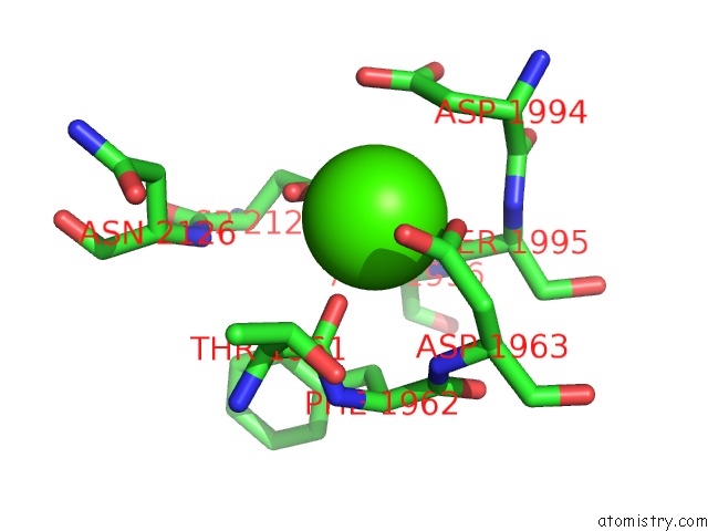
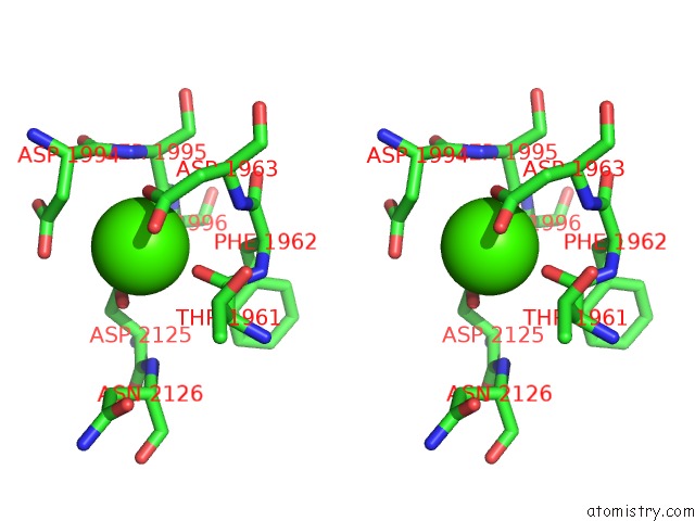
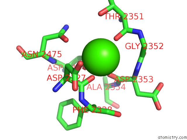
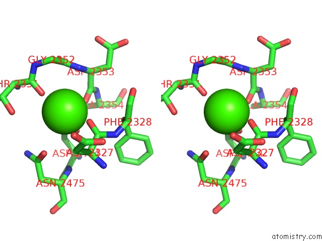
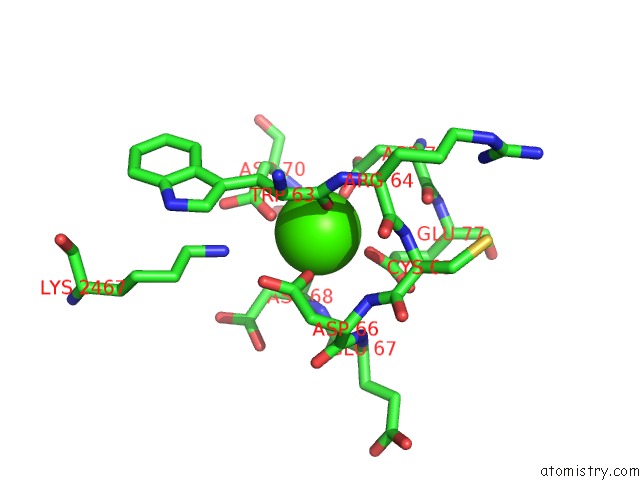
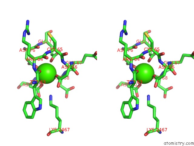
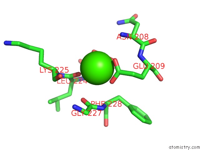
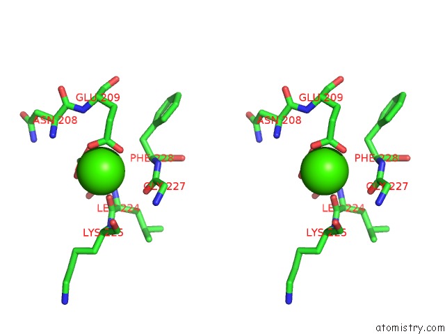
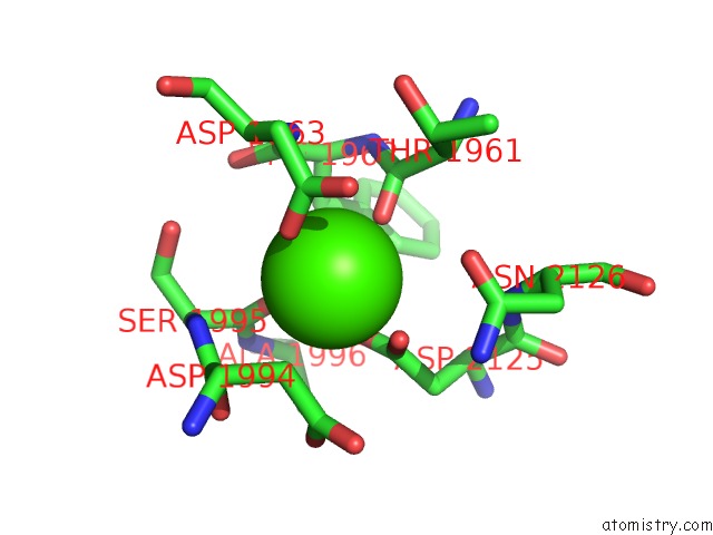
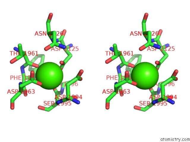
Calcium binding site 1 out of 16 in 5b4x

Go back to Calcium Binding Sites List in 5b4x
Calcium binding site 1 out of 16 in the Crystal Structure of the APOER2 Ectodomain in Complex with the Reelin R56 Fragment


Mono view


Stereo pair view

A full contact list of Calcium with other atoms in the Ca binding site number 1 of Crystal Structure of the APOER2 Ectodomain in Complex with the Reelin R56 Fragment within 5.0Å range:
probe atom residue distance (Å) B Occ
A:Ca4001

b:0.1
occ:1.00
OD2 A:ASP2125 2.3 0.4 1.0
OD1 A:ASP1963 2.3 1.0 1.0
O A:THR1961 2.3 1.0 1.0
OD1 A:ASP2125 2.3 0.4 1.0
O A:SER1995 2.3 0.3 1.0
CG A:ASP2125 2.7 0.4 1.0
CG A:ASP1963 3.0 0.1 1.0
OD2 A:ASP1963 3.1 0.8 1.0
CB A:ASP1994 3.4 0.1 1.0
C A:SER1995 3.5 0.9 1.0
C A:THR1961 3.5 0.0 1.0
OD2 A:ASP1994 3.6 98.8 1.0
N A:SER1995 3.6 0.4 1.0
CG A:ASP1994 3.7 0.9 1.0
N A:ASP1963 4.0 0.8 1.0
C A:ASP1994 4.2 0.3 1.0
CA A:SER1995 4.2 0.8 1.0
CB A:ASP2125 4.2 0.1 1.0
CB A:ASP1963 4.2 0.7 1.0
CA A:ASP1994 4.3 0.4 1.0
CA A:THR1961 4.3 0.0 1.0
C A:PHE1962 4.4 0.6 1.0
CA A:ASP1963 4.5 0.3 1.0
CB A:THR1961 4.5 0.9 1.0
N A:PHE1962 4.5 0.2 1.0
CB A:PHE1962 4.5 0.2 1.0
OD1 A:ASP1994 4.5 0.7 1.0
N A:ALA1996 4.6 0.2 1.0
N A:ASN2126 4.7 92.5 1.0
CA A:PHE1962 4.7 0.7 1.0
CA A:ALA1996 4.9 0.8 1.0
CA A:ASP2125 5.0 93.9 1.0
CB A:SER1995 5.0 0.8 1.0

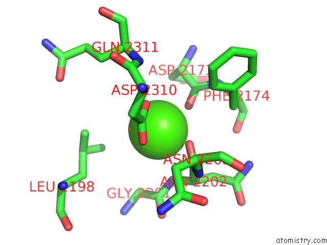
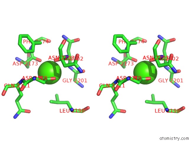
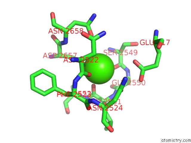
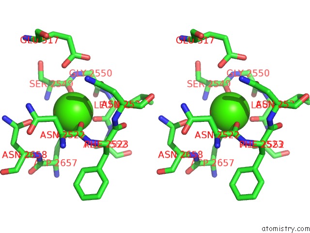
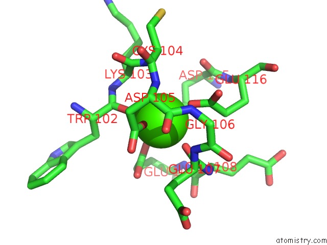
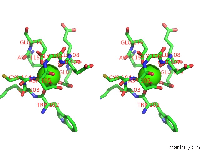
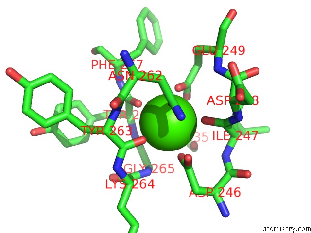
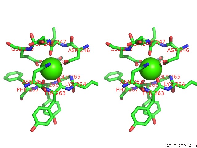
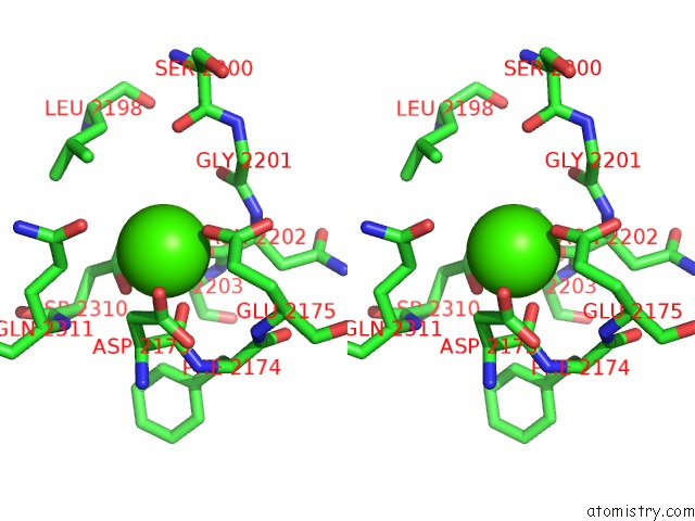
Calcium binding site 2 out of 16 in 5b4x

Go back to Calcium Binding Sites List in 5b4x
Calcium binding site 2 out of 16 in the Crystal Structure of the APOER2 Ectodomain in Complex with the Reelin R56 Fragment


Mono view


Stereo pair view

A full contact list of Calcium with other atoms in the Ca binding site number 2 of Crystal Structure of the APOER2 Ectodomain in Complex with the Reelin R56 Fragment within 5.0Å range:
probe atom residue distance (Å) B Occ
A:Ca4002

b:0.1
occ:1.00
O A:ASN2202 2.3 0.2 1.0
OD1 A:ASP2310 2.3 0.7 1.0
OD2 A:ASP2310 2.3 1.0 1.0
O A:ASP2173 2.3 0.2 1.0
CG A:ASP2310 2.7 0.6 1.0
N A:ASN2202 3.2 0.6 1.0
C A:ASN2202 3.2 0.6 1.0
C A:ASP2173 3.5 0.8 1.0
CA A:ASN2202 3.7 1.0 1.0
CD2 A:LEU2198 4.0 0.9 1.0
CA A:ASP2173 4.1 0.2 1.0
C A:GLY2201 4.1 0.3 1.0
CB A:ASP2173 4.2 0.5 1.0
CB A:ASP2310 4.2 0.2 1.0
N A:ASN2203 4.3 0.1 1.0
CA A:GLY2201 4.3 0.9 1.0
CD1 A:LEU2198 4.4 0.7 1.0
CB A:ASN2202 4.4 0.3 1.0
CA A:ASN2203 4.6 0.2 1.0
N A:PHE2174 4.6 0.1 1.0
CG A:LEU2198 4.8 0.5 1.0
CB A:PHE2174 4.8 1.0 1.0
N A:GLN2311 4.8 96.6 1.0
CA A:ASP2310 5.0 0.1 1.0

Calcium binding site 3 out of 16 in 5b4x

Go back to Calcium Binding Sites List in 5b4x
Calcium binding site 3 out of 16 in the Crystal Structure of the APOER2 Ectodomain in Complex with the Reelin R56 Fragment


Mono view


Stereo pair view

A full contact list of Calcium with other atoms in the Ca binding site number 3 of Crystal Structure of the APOER2 Ectodomain in Complex with the Reelin R56 Fragment within 5.0Å range:
probe atom residue distance (Å) B Occ
A:Ca4003

b:66.3
occ:1.00
O A:ASP2353 2.3 65.9 1.0
O A:ASP2327 2.3 65.5 1.0
OD2 A:ASP2474 2.3 68.4 1.0
OD1 A:ASP2474 2.3 72.6 1.0
O A:THR2351 2.3 71.4 1.0
CG A:ASP2474 2.7 68.0 1.0
C A:ASP2353 3.5 72.5 1.0
C A:ASP2327 3.5 66.3 1.0
C A:THR2351 3.6 65.6 1.0
N A:ASP2353 3.7 72.9 1.0
OD1 A:ASN2475 3.7 65.1 1.0
C A:GLY2352 4.0 66.0 1.0
CA A:ASP2327 4.1 69.0 1.0
CA A:ASP2353 4.1 75.2 1.0
OG1 A:THR2351 4.1 66.5 1.0
CA A:GLY2352 4.2 63.7 1.0
CB A:ASP2474 4.2 65.1 1.0
CB A:ASP2327 4.3 70.7 1.0
N A:GLY2352 4.3 62.5 1.0
CG A:ASN2475 4.5 64.6 1.0
N A:ALA2354 4.5 71.0 1.0
N A:PHE2328 4.6 65.0 1.0
O A:GLY2352 4.6 64.9 1.0
ND2 A:ASN2475 4.7 67.9 1.0
CA A:THR2351 4.7 66.1 1.0
CB A:PHE2328 4.8 69.4 1.0
N A:ASN2475 5.0 61.3 1.0
N A:THR2351 5.0 68.7 1.0
CA A:ALA2354 5.0 70.7 1.0

Calcium binding site 4 out of 16 in 5b4x

Go back to Calcium Binding Sites List in 5b4x
Calcium binding site 4 out of 16 in the Crystal Structure of the APOER2 Ectodomain in Complex with the Reelin R56 Fragment


Mono view


Stereo pair view

A full contact list of Calcium with other atoms in the Ca binding site number 4 of Crystal Structure of the APOER2 Ectodomain in Complex with the Reelin R56 Fragment within 5.0Å range:
probe atom residue distance (Å) B Occ
A:Ca4004

b:62.9
occ:1.00
O A:SER2549 2.3 70.0 1.0
O A:LEU2551 2.3 56.9 1.0
OD2 A:ASP2657 2.3 66.0 1.0
O A:ASN2522 2.3 62.1 1.0
OD1 A:ASP2657 2.3 64.7 1.0
CG A:ASP2657 2.7 63.3 1.0
C A:ASN2522 3.5 58.0 1.0
C A:SER2549 3.5 71.1 1.0
C A:LEU2551 3.5 58.0 1.0
OD1 A:ASN2658 4.0 85.2 1.0
N A:LEU2551 4.0 63.2 1.0
CA A:ASN2522 4.1 59.8 1.0
CB A:ASN2522 4.2 67.1 1.0
N A:SER2549 4.2 72.5 1.0
CB A:ASP2657 4.2 57.9 1.0
C A:GLY2550 4.3 64.8 1.0
OE1 D:GLU517 4.3 0.0 1.0
CA A:SER2549 4.3 71.4 1.0
CA A:LEU2551 4.3 59.4 1.0
OD1 A:ASN2524 4.5 72.8 1.0
CB A:SER2549 4.5 72.4 1.0
N A:GLY2550 4.6 67.3 1.0
N A:ALA2552 4.6 56.8 1.0
N A:PHE2523 4.6 56.9 1.0
O A:GLY2550 4.6 70.8 1.0
CA A:GLY2550 4.7 65.2 1.0
CA A:ALA2552 4.8 60.1 1.0
N A:ASN2658 4.9 60.7 1.0
C A:PHE2523 4.9 59.9 1.0
CB A:PHE2523 4.9 58.1 1.0
CB A:LEU2551 4.9 61.1 1.0
CA A:PHE2523 5.0 58.9 1.0
O A:PHE2523 5.0 59.1 1.0

Calcium binding site 5 out of 16 in 5b4x

Go back to Calcium Binding Sites List in 5b4x
Calcium binding site 5 out of 16 in the Crystal Structure of the APOER2 Ectodomain in Complex with the Reelin R56 Fragment


Mono view


Stereo pair view

A full contact list of Calcium with other atoms in the Ca binding site number 5 of Crystal Structure of the APOER2 Ectodomain in Complex with the Reelin R56 Fragment within 5.0Å range:
probe atom residue distance (Å) B Occ
B:Ca5001

b:86.3
occ:1.00
O B:ASP68 2.3 97.7 1.0
O B:TRP63 2.3 87.6 1.0
OD2 B:ASP70 2.3 80.0 1.0
OD1 B:ASP66 2.3 92.2 1.0
OE2 B:GLU77 2.3 98.9 1.0
OD2 B:ASP76 2.3 94.1 1.0
C B:ASP68 3.4 0.1 1.0
CD B:GLU77 3.4 0.4 1.0
C B:TRP63 3.5 84.1 1.0
CG B:ASP66 3.5 98.7 1.0
CG B:ASP70 3.5 84.4 1.0
CG B:ASP76 3.6 97.1 1.0
N B:ASP68 3.8 0.0 1.0
CB B:ASP68 3.9 0.4 1.0
CA B:ASP68 3.9 0.8 1.0
NZ A:LYS2467 4.0 61.2 1.0
OD2 B:ASP66 4.0 97.0 1.0
CB B:TRP63 4.1 66.4 1.0
CG B:GLU77 4.1 0.9 1.0
CA B:TRP63 4.1 75.9 1.0
CB B:ASP70 4.2 87.2 1.0
N B:ASP66 4.2 0.9 1.0
N B:ASP70 4.3 0.3 1.0
CB B:ASP76 4.3 95.7 1.0
OE1 B:GLU77 4.4 97.6 1.0
N B:CYS65 4.4 0.2 1.0
OD1 B:ASP76 4.5 97.9 1.0
OD1 B:ASP70 4.5 85.8 1.0
N B:ASP69 4.5 0.5 1.0
N B:ARG64 4.5 91.7 1.0
N B:GLU67 4.6 0.2 1.0
CB B:ASP66 4.7 0.1 1.0
O B:ASP76 4.7 96.7 1.0
CA B:ASP66 4.8 0.9 1.0
C B:ARG64 4.8 0.3 1.0
CA B:ARG64 4.8 99.3 1.0
C B:ASP66 4.8 0.8 1.0
CA B:ASP69 4.9 0.6 1.0
CA B:ASP70 4.9 93.2 1.0
C B:GLU67 5.0 0.9 1.0
C B:ASP69 5.0 0.3 1.0

Calcium binding site 6 out of 16 in 5b4x

Go back to Calcium Binding Sites List in 5b4x
Calcium binding site 6 out of 16 in the Crystal Structure of the APOER2 Ectodomain in Complex with the Reelin R56 Fragment


Mono view


Stereo pair view

A full contact list of Calcium with other atoms in the Ca binding site number 6 of Crystal Structure of the APOER2 Ectodomain in Complex with the Reelin R56 Fragment within 5.0Å range:
probe atom residue distance (Å) B Occ
B:Ca5002

b:0.3
occ:1.00
O B:TRP102 2.3 0.4 1.0
O B:GLU107 2.3 0.6 1.0
OD2 B:ASP115 2.3 0.9 1.0
OE2 B:GLU116 2.3 0.5 1.0
OE1 B:GLU109 2.3 0.8 1.0
OD2 B:ASP105 2.3 0.7 1.0
C B:TRP102 3.3 0.7 1.0
CD B:GLU109 3.5 0.7 1.0
CD B:GLU116 3.5 0.3 1.0
C B:GLU107 3.5 0.6 1.0
CG B:ASP115 3.6 0.4 1.0
CG B:ASP105 3.6 0.7 1.0
CA B:LYS103 3.9 0.2 1.0
N B:CYS104 3.9 0.4 1.0
OE2 B:GLU109 3.9 0.5 1.0
N B:LYS103 4.0 0.3 1.0
N B:GLU107 4.1 0.6 1.0
N B:ASP105 4.1 0.4 1.0
CG B:GLU116 4.2 0.6 1.0
C B:LYS103 4.2 0.3 1.0
CA B:GLU107 4.3 0.1 1.0
OD1 B:ASP115 4.3 0.7 1.0
OD1 B:ASP105 4.3 0.9 1.0
OE1 B:GLU116 4.5 0.1 1.0
CA B:TRP102 4.5 0.2 1.0
N B:GLU108 4.5 0.6 1.0
N B:GLU109 4.6 0.6 1.0
CB B:TRP102 4.6 0.9 1.0
CB B:ASP105 4.6 0.8 1.0
CB B:ASP115 4.6 0.4 1.0
CB B:GLU107 4.6 0.8 1.0
CG B:GLU109 4.7 0.9 1.0
CA B:GLU108 4.7 0.5 1.0
CA B:ASP105 4.8 0.1 1.0
N B:GLY106 4.8 0.6 1.0
CB B:GLU109 4.9 1.0 1.0
CA B:CYS104 4.9 0.3 1.0
O B:ASP115 5.0 0.2 1.0
C B:ASP105 5.0 1.0 1.0
C B:GLU108 5.0 0.7 1.0

Calcium binding site 7 out of 16 in 5b4x

Go back to Calcium Binding Sites List in 5b4x
Calcium binding site 7 out of 16 in the Crystal Structure of the APOER2 Ectodomain in Complex with the Reelin R56 Fragment


Mono view


Stereo pair view

A full contact list of Calcium with other atoms in the Ca binding site number 7 of Crystal Structure of the APOER2 Ectodomain in Complex with the Reelin R56 Fragment within 5.0Å range:
probe atom residue distance (Å) B Occ
B:Ca5003

b:0.3
occ:1.00
OD2 B:ASP223 2.3 0.3 1.0
OE1 B:GLU209 2.3 0.2 1.0
OE2 B:GLU209 2.3 0.6 1.0
OD1 B:ASP223 2.3 0.8 1.0
O B:LEU224 2.3 1.0 1.0
CG B:ASP223 2.6 0.5 1.0
CD B:GLU209 2.6 0.6 1.0
C B:LEU224 3.6 0.8 1.0
O B:GLY227 3.8 0.0 1.0
CB B:ASP223 4.0 0.9 1.0
N B:LEU224 4.0 0.9 1.0
C B:GLY227 4.1 0.3 1.0
CG B:GLU209 4.2 0.9 1.0
C B:ASP223 4.3 0.3 1.0
CA B:ASP223 4.3 0.7 1.0
N B:PHE228 4.4 0.2 1.0
O B:LYS225 4.4 0.6 1.0
CB B:PHE228 4.4 0.4 1.0
N B:ASN208 4.5 0.9 1.0
CA B:LEU224 4.5 0.3 1.0
CA B:PHE228 4.5 0.5 1.0
N B:LYS225 4.5 0.5 1.0
CA B:LYS225 4.5 0.5 1.0
C B:LYS225 4.7 0.9 1.0
N B:GLU209 4.7 0.5 1.0
CB B:GLU209 4.9 0.2 1.0
N B:GLY227 4.9 0.7 1.0
CA B:GLY227 4.9 0.6 1.0

Calcium binding site 8 out of 16 in 5b4x

Go back to Calcium Binding Sites List in 5b4x
Calcium binding site 8 out of 16 in the Crystal Structure of the APOER2 Ectodomain in Complex with the Reelin R56 Fragment


Mono view


Stereo pair view

A full contact list of Calcium with other atoms in the Ca binding site number 8 of Crystal Structure of the APOER2 Ectodomain in Complex with the Reelin R56 Fragment within 5.0Å range:
probe atom residue distance (Å) B Occ
B:Ca5004

b:0.1
occ:1.00
OE1 B:GLU249 2.3 0.3 1.0
OD1 B:ASP246 2.3 0.8 1.0
O B:TYR263 2.3 0.5 1.0
OD1 B:ASN262 2.3 0.9 1.0
O B:ILE247 2.3 0.8 1.0
O B:TYR266 2.3 0.8 1.0
CG B:ASP246 3.1 0.7 1.0
CD B:GLU249 3.1 0.5 1.0
OD2 B:ASP246 3.2 0.1 1.0
OE2 B:GLU249 3.3 0.7 1.0
C B:TYR266 3.4 0.8 1.0
N B:TYR266 3.4 0.0 1.0
CG B:ASN262 3.5 0.1 1.0
C B:TYR263 3.5 0.4 1.0
C B:ILE247 3.5 0.4 1.0
N B:GLY265 3.6 0.4 1.0
CA B:TYR266 3.9 0.3 1.0
O B:GLY235 3.9 0.2 1.0
N B:TYR263 4.0 0.1 1.0
ND2 B:ASN262 4.1 0.9 1.0
C B:GLY265 4.1 0.8 1.0
CA B:GLY265 4.2 0.1 1.0
N B:ILE247 4.3 0.1 1.0
C B:LYS264 4.3 0.5 1.0
CA B:ILE247 4.4 0.2 1.0
CA B:TYR263 4.4 0.6 1.0
N B:LYS264 4.4 0.5 1.0
N B:PHE267 4.5 94.4 1.0
CA B:LYS264 4.5 0.4 1.0
N B:ASP248 4.5 0.9 1.0
CG B:GLU249 4.6 0.3 1.0
CB B:ASP246 4.6 0.5 1.0
CB B:ILE247 4.6 0.4 1.0
N B:GLU249 4.6 0.1 1.0
CB B:ASN262 4.7 99.7 1.0
C B:ASN262 4.8 0.1 1.0
CA B:ASP248 4.8 0.6 1.0
CA B:ASN262 4.8 97.2 1.0
CB B:PHE267 4.9 91.3 1.0
C B:ASP246 5.0 0.5 1.0

Calcium binding site 9 out of 16 in 5b4x

Go back to Calcium Binding Sites List in 5b4x
Calcium binding site 9 out of 16 in the Crystal Structure of the APOER2 Ectodomain in Complex with the Reelin R56 Fragment


Mono view


Stereo pair view

A full contact list of Calcium with other atoms in the Ca binding site number 9 of Crystal Structure of the APOER2 Ectodomain in Complex with the Reelin R56 Fragment within 5.0Å range:
probe atom residue distance (Å) B Occ
C:Ca4001

b:0.2
occ:1.00
OD1 C:ASP1963 2.3 0.3 1.0
OD2 C:ASP2125 2.3 0.7 1.0
O C:THR1961 2.3 1.0 1.0
OD1 C:ASP2125 2.3 0.1 1.0
O C:SER1995 2.3 0.7 1.0
CG C:ASP2125 2.7 0.4 1.0
CG C:ASP1963 3.2 0.6 1.0
CB C:ASP1994 3.4 0.7 1.0
C C:SER1995 3.4 0.0 1.0
C C:THR1961 3.5 0.9 1.0
N C:SER1995 3.6 0.9 1.0
OD2 C:ASP1963 3.8 0.8 1.0
C C:ASP1994 3.8 0.5 1.0
N C:ASP1963 3.9 0.3 1.0
CG C:ASP1994 3.9 0.2 1.0
C C:PHE1962 4.0 0.9 1.0
OD1 C:ASN2126 4.0 1.0 1.0
CA C:ASP1994 4.0 0.0 1.0
OD1 C:ASP1994 4.0 97.7 1.0
CB C:PHE1962 4.1 0.1 1.0
CA C:SER1995 4.1 0.4 1.0
CB C:ASP2125 4.2 0.8 1.0
CA C:ASP1963 4.2 0.0 1.0
CB C:ASP1963 4.3 0.3 1.0
N C:PHE1962 4.3 0.3 1.0
CA C:PHE1962 4.3 0.6 1.0
O C:ASP1994 4.4 0.4 1.0
CA C:THR1961 4.5 0.8 1.0
O C:PHE1962 4.5 0.6 1.0
N C:ALA1996 4.5 0.8 1.0
CB C:THR1961 4.5 0.6 1.0
OG C:SER1995 4.5 0.4 1.0
CG C:ASN2126 4.6 0.9 1.0
N C:ASN2126 4.7 98.3 1.0
CD2 C:PHE1962 4.7 0.1 1.0
ND2 C:ASN2126 4.7 0.6 1.0
OD2 C:ASP1994 4.8 0.2 1.0
CA C:ALA1996 4.8 0.8 1.0
CA C:ASP2125 4.9 95.4 1.0
CG C:PHE1962 4.9 0.7 1.0
CB C:SER1995 4.9 0.7 1.0

Calcium binding site 10 out of 16 in 5b4x

Go back to Calcium Binding Sites List in 5b4x
Calcium binding site 10 out of 16 in the Crystal Structure of the APOER2 Ectodomain in Complex with the Reelin R56 Fragment


Mono view


Stereo pair view

A full contact list of Calcium with other atoms in the Ca binding site number 10 of Crystal Structure of the APOER2 Ectodomain in Complex with the Reelin R56 Fragment within 5.0Å range:
probe atom residue distance (Å) B Occ
C:Ca4002

b:0.5
occ:1.00
OE2 C:GLU2175 2.3 0.2 1.0
OD1 C:ASP2310 2.3 0.7 1.0
O C:ASN2202 2.3 0.2 1.0
OD2 C:ASP2310 2.3 0.3 1.0
O C:ASP2173 2.3 0.3 1.0
CG C:ASP2310 2.7 0.9 1.0
C C:ASP2173 3.4 0.4 1.0
CD C:GLU2175 3.4 0.9 1.0
C C:ASN2202 3.5 0.9 1.0
CB C:ASP2173 3.8 0.8 1.0
CA C:ASP2173 4.0 0.0 1.0
N C:ASN2202 4.1 0.9 1.0
CG C:GLN2311 4.1 0.8 1.0
C C:GLY2201 4.1 0.2 1.0
CG C:GLU2175 4.1 0.9 1.0
CB C:ASP2310 4.2 0.8 1.0
CA C:ASN2202 4.3 0.4 1.0
OE1 C:GLU2175 4.4 0.6 1.0
O C:GLY2201 4.4 0.8 1.0
OE1 C:GLN2311 4.4 0.7 1.0
CG C:ASP2173 4.5 0.4 1.0
N C:ASN2203 4.5 0.8 1.0
N C:PHE2174 4.6 1.0 1.0
CA C:GLY2201 4.6 0.7 1.0
CD C:GLN2311 4.7 0.0 1.0
CA C:ASN2203 4.7 0.2 1.0
CD2 C:LEU2198 4.7 0.3 1.0
N C:GLN2311 4.8 93.1 1.0
O C:SER2200 4.8 0.8 1.0
OD2 C:ASP2173 4.9 0.8 1.0
N C:GLU2175 4.9 0.1 1.0

Reference:

H.Hirai, N.Yasui, K.Yamashita, S.Tabata, M.Yamamoto, J.Takagi, T.Nogi. Crystal Structure of the Ectodomain From A Ldlr Close Homologue in Complex with Its Physiological Ligand. To Be Published.
Page generated: Sat Dec 12 05:18:13 2020

Last articles

Zn in 8WB0
Zn in 8WAX
Zn in 8WAU
Zn in 8WAZ
Zn in 8WAY
Zn in 8WAV
Zn in 8WAW
Zn in 8WAT
Zn in 8W7M
Zn in 8WD3
© Copyright 2008-2020 by atomistry.com
Home   |    Site Map   |    Copyright   |    Contact us   |    Privacy