Calcium »
PDB 5b5e-5byn »
5b5s »
Calcium in PDB 5b5s: Crystal Structure of A Carbohydrate Esterase Family 3 From Talaromyces Cellulolyticus
Protein crystallography data
The structure of Crystal Structure of A Carbohydrate Esterase Family 3 From Talaromyces Cellulolyticus, PDB code: 5b5s
was solved by
M.Watanabe,
K.Ishikawa,
with X-Ray Crystallography technique. A brief refinement statistics is given in the table below:
| Resolution Low / High (Å) | 30.00 / 1.50 |
| Space group | P 41 21 2 |
| Cell size a, b, c (Å), α, β, γ (°) | 70.905, 70.905, 87.093, 90.00, 90.00, 90.00 |
| R / Rfree (%) | 14.5 / 20.3 |
Calcium Binding Sites:
The binding sites of Calcium atom in the Crystal Structure of A Carbohydrate Esterase Family 3 From Talaromyces Cellulolyticus
(pdb code 5b5s). This binding sites where shown within
5.0 Angstroms radius around Calcium atom.
In total only one binding site of Calcium was determined in the Crystal Structure of A Carbohydrate Esterase Family 3 From Talaromyces Cellulolyticus, PDB code: 5b5s:
In total only one binding site of Calcium was determined in the Crystal Structure of A Carbohydrate Esterase Family 3 From Talaromyces Cellulolyticus, PDB code: 5b5s:
Calcium binding site 1 out of 1 in 5b5s
Go back to
Calcium binding site 1 out
of 1 in the Crystal Structure of A Carbohydrate Esterase Family 3 From Talaromyces Cellulolyticus
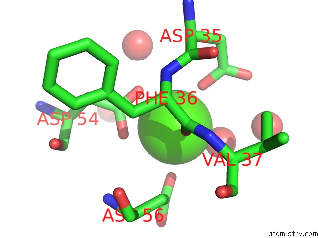
Mono view
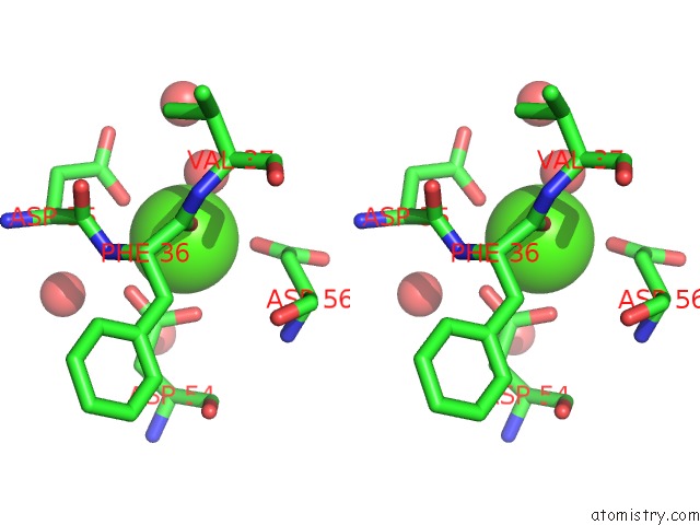
Stereo pair view
Mono view
Stereo pair view
A full contact list of Calcium with other atoms in the Ca binding
site number 1 of Crystal Structure of A Carbohydrate Esterase Family 3 From Talaromyces Cellulolyticus within 5.0Å range:
|
Reference:
M.Watanabe,
H.Fukada,
H.Inoue,
K.Ishikawa.
Crystal Structure of An Acetylesterase From Talaromyces Cellulolyticus and the Importance of A Disulfide Bond Near the Active Site Febs Lett. V. 589 1200 2015.
ISSN: ISSN 0014-5793
PubMed: 25825334
DOI: 10.1016/J.FEBSLET.2015.03.020
Page generated: Sun Jul 14 16:49:57 2024
ISSN: ISSN 0014-5793
PubMed: 25825334
DOI: 10.1016/J.FEBSLET.2015.03.020
Last articles
Zn in 9J0NZn in 9J0O
Zn in 9J0P
Zn in 9FJX
Zn in 9EKB
Zn in 9C0F
Zn in 9CAH
Zn in 9CH0
Zn in 9CH3
Zn in 9CH1