Calcium »
PDB 5b5e-5byn »
5bqt »
Calcium in PDB 5bqt: Structure of TRMBL2, An Archaeal Chromatin Protein, Shows A Novel Mode of Dna Binding.
Protein crystallography data
The structure of Structure of TRMBL2, An Archaeal Chromatin Protein, Shows A Novel Mode of Dna Binding., PDB code: 5bqt
was solved by
M.U.Ahmad,
K.Diederichs,
W.Welte,
with X-Ray Crystallography technique. A brief refinement statistics is given in the table below:
| Resolution Low / High (Å) | 47.84 / 3.00 |
| Space group | P 21 21 2 |
| Cell size a, b, c (Å), α, β, γ (°) | 82.298, 235.143, 63.511, 90.00, 90.00, 90.00 |
| R / Rfree (%) | 23.3 / 28.9 |
Calcium Binding Sites:
The binding sites of Calcium atom in the Structure of TRMBL2, An Archaeal Chromatin Protein, Shows A Novel Mode of Dna Binding.
(pdb code 5bqt). This binding sites where shown within
5.0 Angstroms radius around Calcium atom.
In total 2 binding sites of Calcium where determined in the Structure of TRMBL2, An Archaeal Chromatin Protein, Shows A Novel Mode of Dna Binding., PDB code: 5bqt:
Jump to Calcium binding site number: 1; 2;
In total 2 binding sites of Calcium where determined in the Structure of TRMBL2, An Archaeal Chromatin Protein, Shows A Novel Mode of Dna Binding., PDB code: 5bqt:
Jump to Calcium binding site number: 1; 2;
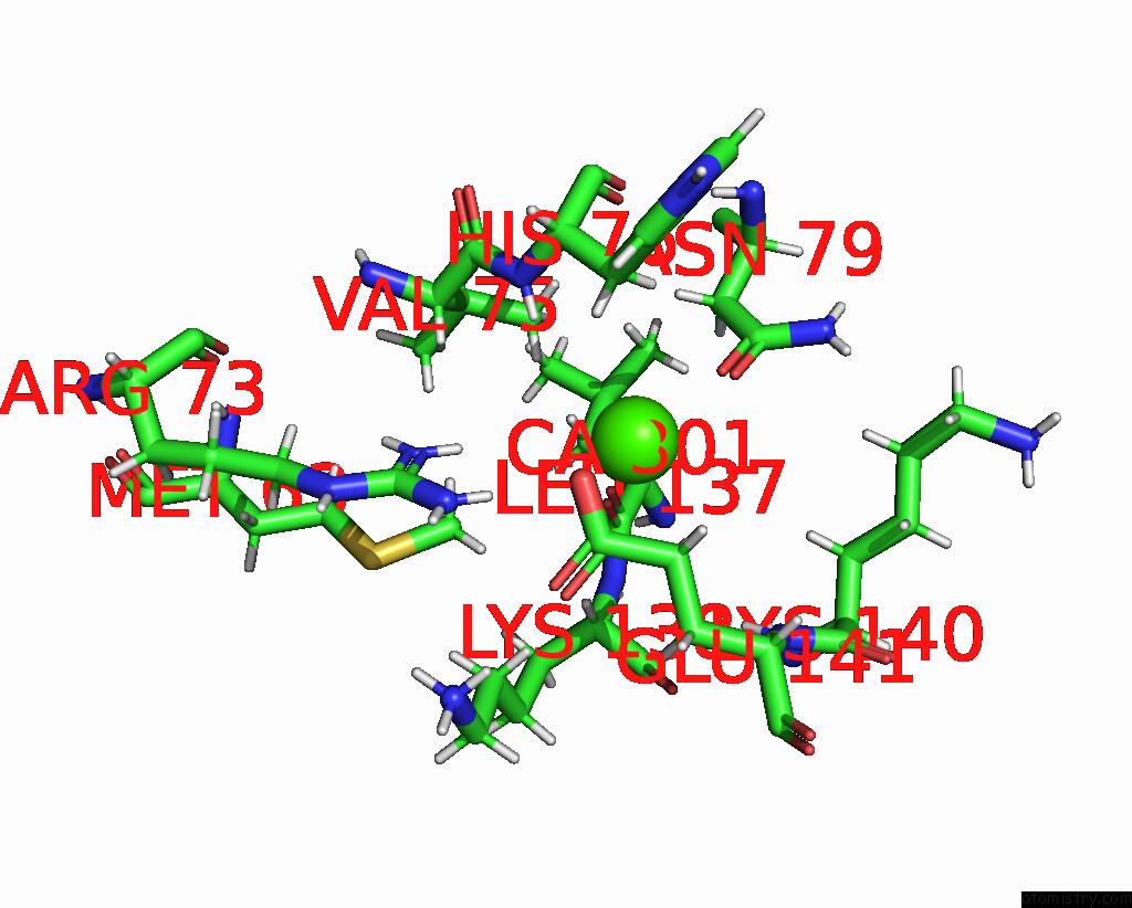
Calcium binding site 1 out of 2 in 5bqt
Go back to
Calcium binding site 1 out
of 2 in the Structure of TRMBL2, An Archaeal Chromatin Protein, Shows A Novel Mode of Dna Binding.
Mono view

Stereo pair view
Mono view
Stereo pair view
A full contact list of Calcium with other atoms in the Ca binding
site number 1 of Structure of TRMBL2, An Archaeal Chromatin Protein, Shows A Novel Mode of Dna Binding. within 5.0Å range:
|
Calcium binding site 2 out of 2 in 5bqt
Go back to
Calcium binding site 2 out
of 2 in the Structure of TRMBL2, An Archaeal Chromatin Protein, Shows A Novel Mode of Dna Binding.

Mono view

Stereo pair view
Mono view
Stereo pair view
A full contact list of Calcium with other atoms in the Ca binding
site number 2 of Structure of TRMBL2, An Archaeal Chromatin Protein, Shows A Novel Mode of Dna Binding. within 5.0Å range:
|
Reference:
M.U.Ahmad,
I.Waege,
W.Hausner,
M.Thomm,
W.Boos,
K.Diederichs,
W.Welte.
Structural Insights Into Nonspecific Binding of Dna By TRMBL2, An Archaeal Chromatin Protein. J.Mol.Biol. V. 427 3216 2015.
ISSN: ESSN 1089-8638
PubMed: 26299937
DOI: 10.1016/J.JMB.2015.08.012
Page generated: Wed Jul 9 04:26:39 2025
ISSN: ESSN 1089-8638
PubMed: 26299937
DOI: 10.1016/J.JMB.2015.08.012
Last articles
Fe in 2YXOFe in 2YRS
Fe in 2YXC
Fe in 2YNM
Fe in 2YVJ
Fe in 2YP1
Fe in 2YU2
Fe in 2YU1
Fe in 2YQB
Fe in 2YOO