Calcium »
PDB 5b5e-5byn »
5bwg »
Calcium in PDB 5bwg: Structure of H200C Variant of Homoprotocatechuate 2,3-Dioxygenase From B.Fuscum at 1.75 Ang Resolution
Protein crystallography data
The structure of Structure of H200C Variant of Homoprotocatechuate 2,3-Dioxygenase From B.Fuscum at 1.75 Ang Resolution, PDB code: 5bwg
was solved by
E.G.Kovaleva,
J.D.Lipscomb,
with X-Ray Crystallography technique. A brief refinement statistics is given in the table below:
| Resolution Low / High (Å) | 45.73 / 1.75 |
| Space group | P 21 21 2 |
| Cell size a, b, c (Å), α, β, γ (°) | 110.748, 150.627, 96.269, 90.00, 90.00, 90.00 |
| R / Rfree (%) | 14.5 / 17.9 |
Other elements in 5bwg:
The structure of Structure of H200C Variant of Homoprotocatechuate 2,3-Dioxygenase From B.Fuscum at 1.75 Ang Resolution also contains other interesting chemical elements:
| Iron | (Fe) | 4 atoms |
| Chlorine | (Cl) | 4 atoms |
Calcium Binding Sites:
The binding sites of Calcium atom in the Structure of H200C Variant of Homoprotocatechuate 2,3-Dioxygenase From B.Fuscum at 1.75 Ang Resolution
(pdb code 5bwg). This binding sites where shown within
5.0 Angstroms radius around Calcium atom.
In total only one binding site of Calcium was determined in the Structure of H200C Variant of Homoprotocatechuate 2,3-Dioxygenase From B.Fuscum at 1.75 Ang Resolution, PDB code: 5bwg:
In total only one binding site of Calcium was determined in the Structure of H200C Variant of Homoprotocatechuate 2,3-Dioxygenase From B.Fuscum at 1.75 Ang Resolution, PDB code: 5bwg:
Calcium binding site 1 out of 1 in 5bwg
Go back to
Calcium binding site 1 out
of 1 in the Structure of H200C Variant of Homoprotocatechuate 2,3-Dioxygenase From B.Fuscum at 1.75 Ang Resolution
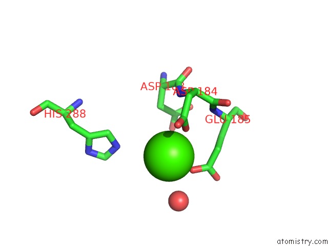
Mono view
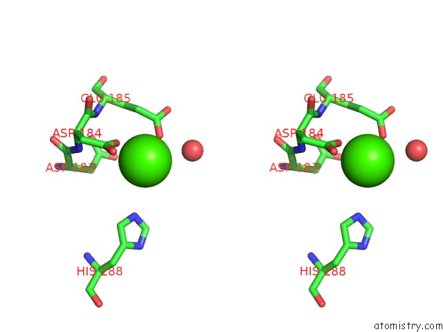
Stereo pair view
Mono view
Stereo pair view
A full contact list of Calcium with other atoms in the Ca binding
site number 1 of Structure of H200C Variant of Homoprotocatechuate 2,3-Dioxygenase From B.Fuscum at 1.75 Ang Resolution within 5.0Å range:
|
Reference:
K.K.Meier,
M.S.Rogers,
E.G.Kovaleva,
M.M.Mbughuni,
E.L.Bominaar,
J.D.Lipscomb,
E.Munck.
A Long-Lived Fe(III)-(Hydroperoxo) Intermediate in the Active H200C Variant of Homoprotocatechuate 2,3-Dioxygenase: Characterization By Mossbauer, Electron Paramagnetic Resonance, and Density Functional Theory Methods. Inorg.Chem. V. 54 10269 2015.
ISSN: ISSN 0020-1669
PubMed: 26485328
DOI: 10.1021/ACS.INORGCHEM.5B01576
Page generated: Wed Jul 9 04:28:36 2025
ISSN: ISSN 0020-1669
PubMed: 26485328
DOI: 10.1021/ACS.INORGCHEM.5B01576
Last articles
Fe in 2YXOFe in 2YRS
Fe in 2YXC
Fe in 2YNM
Fe in 2YVJ
Fe in 2YP1
Fe in 2YU2
Fe in 2YU1
Fe in 2YQB
Fe in 2YOO