Calcium »
PDB 5ckm-5cxl »
5ckq »
Calcium in PDB 5ckq: CUB1-Egf-CUB2 Domains of Rat Masp-1
Protein crystallography data
The structure of CUB1-Egf-CUB2 Domains of Rat Masp-1, PDB code: 5ckq
was solved by
R.Nan,
C.M.Furze,
D.W.Wright,
J.Gor,
R.Wallis,
S.J.Perkins,
with X-Ray Crystallography technique. A brief refinement statistics is given in the table below:
| Resolution Low / High (Å) | 76.36 / 3.70 |
| Space group | I 2 3 |
| Cell size a, b, c (Å), α, β, γ (°) | 152.710, 152.710, 152.710, 90.00, 90.00, 90.00 |
| R / Rfree (%) | 24.8 / 29.4 |
Other elements in 5ckq:
The structure of CUB1-Egf-CUB2 Domains of Rat Masp-1 also contains other interesting chemical elements:
| Sodium | (Na) | 1 atom |
Calcium Binding Sites:
The binding sites of Calcium atom in the CUB1-Egf-CUB2 Domains of Rat Masp-1
(pdb code 5ckq). This binding sites where shown within
5.0 Angstroms radius around Calcium atom.
In total 3 binding sites of Calcium where determined in the CUB1-Egf-CUB2 Domains of Rat Masp-1, PDB code: 5ckq:
Jump to Calcium binding site number: 1; 2; 3;
In total 3 binding sites of Calcium where determined in the CUB1-Egf-CUB2 Domains of Rat Masp-1, PDB code: 5ckq:
Jump to Calcium binding site number: 1; 2; 3;
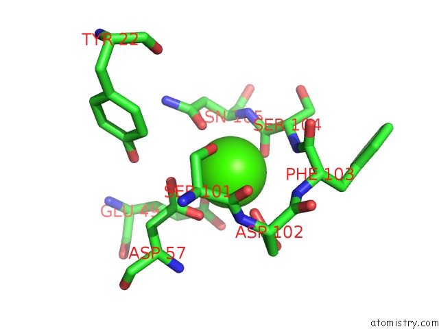
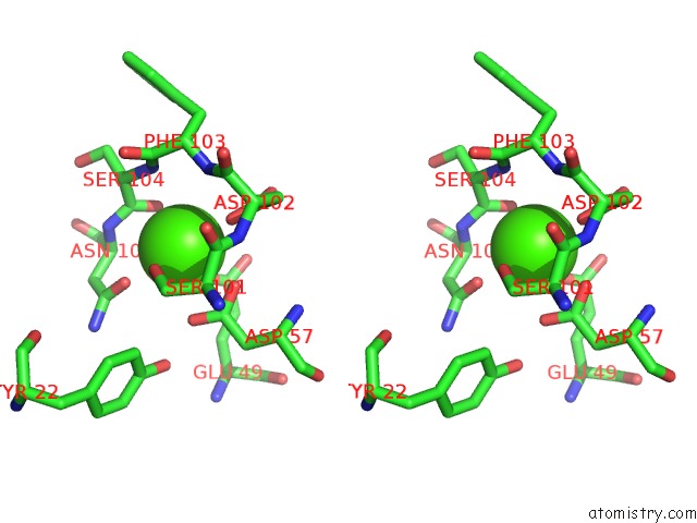
Calcium binding site 1 out of 3 in 5ckq
Go back to
Calcium binding site 1 out
of 3 in the CUB1-Egf-CUB2 Domains of Rat Masp-1
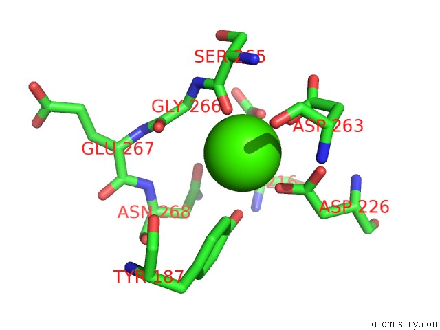
Mono view
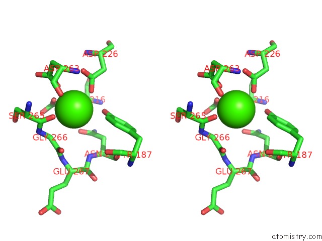
Stereo pair view
Mono view
Stereo pair view
A full contact list of Calcium with other atoms in the Ca binding
site number 1 of CUB1-Egf-CUB2 Domains of Rat Masp-1 within 5.0Å range:
|
Calcium binding site 2 out of 3 in 5ckq
Go back to
Calcium binding site 2 out
of 3 in the CUB1-Egf-CUB2 Domains of Rat Masp-1
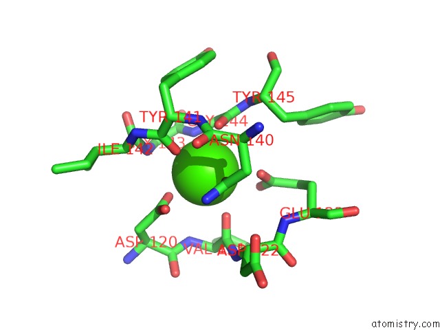
Mono view
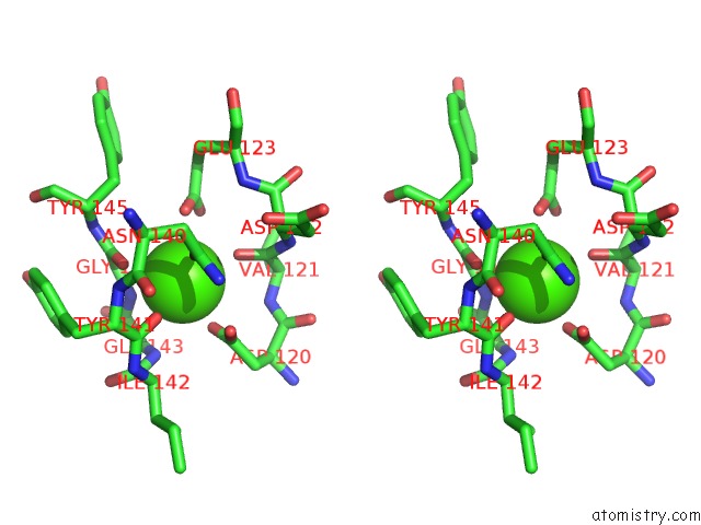
Stereo pair view
Mono view
Stereo pair view
A full contact list of Calcium with other atoms in the Ca binding
site number 2 of CUB1-Egf-CUB2 Domains of Rat Masp-1 within 5.0Å range:
|
Calcium binding site 3 out of 3 in 5ckq
Go back to
Calcium binding site 3 out
of 3 in the CUB1-Egf-CUB2 Domains of Rat Masp-1
Mono view
Stereo pair view
Mono view
Stereo pair view
A full contact list of Calcium with other atoms in the Ca binding
site number 3 of CUB1-Egf-CUB2 Domains of Rat Masp-1 within 5.0Å range:
|
Reference:
R.Nan,
C.M.Furze,
D.W.Wright,
J.Gor,
R.Wallis,
S.J.Perkins.
Flexibility in Mannan-Binding Lectin-Associated Serine Proteases-1 and -2 Provides Insight on Lectin Pathway Activation. Structure V. 25 364 2017.
ISSN: ISSN 1878-4186
PubMed: 28111019
DOI: 10.1016/J.STR.2016.12.014
Page generated: Wed Jul 9 04:36:52 2025
ISSN: ISSN 1878-4186
PubMed: 28111019
DOI: 10.1016/J.STR.2016.12.014
Last articles
Fe in 2YXOFe in 2YRS
Fe in 2YXC
Fe in 2YNM
Fe in 2YVJ
Fe in 2YP1
Fe in 2YU2
Fe in 2YU1
Fe in 2YQB
Fe in 2YOO