Calcium »
PDB 5cy4-5dau »
5d29 »
Calcium in PDB 5d29: X-Ray Structure of Human Glutamate Carboxypeptidase II (Gcpii) in Complex with A Hydroxamate Inhibitor JHU241
Enzymatic activity of X-Ray Structure of Human Glutamate Carboxypeptidase II (Gcpii) in Complex with A Hydroxamate Inhibitor JHU241
All present enzymatic activity of X-Ray Structure of Human Glutamate Carboxypeptidase II (Gcpii) in Complex with A Hydroxamate Inhibitor JHU241:
3.4.17.21;
3.4.17.21;
Protein crystallography data
The structure of X-Ray Structure of Human Glutamate Carboxypeptidase II (Gcpii) in Complex with A Hydroxamate Inhibitor JHU241, PDB code: 5d29
was solved by
C.Barinka,
Z.Novakova,
J.Pavlicek,
with X-Ray Crystallography technique. A brief refinement statistics is given in the table below:
| Resolution Low / High (Å) | 44.82 / 1.80 |
| Space group | I 2 2 2 |
| Cell size a, b, c (Å), α, β, γ (°) | 100.191, 130.459, 157.196, 90.00, 90.00, 90.00 |
| R / Rfree (%) | 17.4 / 20.5 |
Other elements in 5d29:
The structure of X-Ray Structure of Human Glutamate Carboxypeptidase II (Gcpii) in Complex with A Hydroxamate Inhibitor JHU241 also contains other interesting chemical elements:
| Chlorine | (Cl) | 1 atom |
| Zinc | (Zn) | 2 atoms |
Calcium Binding Sites:
The binding sites of Calcium atom in the X-Ray Structure of Human Glutamate Carboxypeptidase II (Gcpii) in Complex with A Hydroxamate Inhibitor JHU241
(pdb code 5d29). This binding sites where shown within
5.0 Angstroms radius around Calcium atom.
In total only one binding site of Calcium was determined in the X-Ray Structure of Human Glutamate Carboxypeptidase II (Gcpii) in Complex with A Hydroxamate Inhibitor JHU241, PDB code: 5d29:
In total only one binding site of Calcium was determined in the X-Ray Structure of Human Glutamate Carboxypeptidase II (Gcpii) in Complex with A Hydroxamate Inhibitor JHU241, PDB code: 5d29:
Calcium binding site 1 out of 1 in 5d29
Go back to
Calcium binding site 1 out
of 1 in the X-Ray Structure of Human Glutamate Carboxypeptidase II (Gcpii) in Complex with A Hydroxamate Inhibitor JHU241
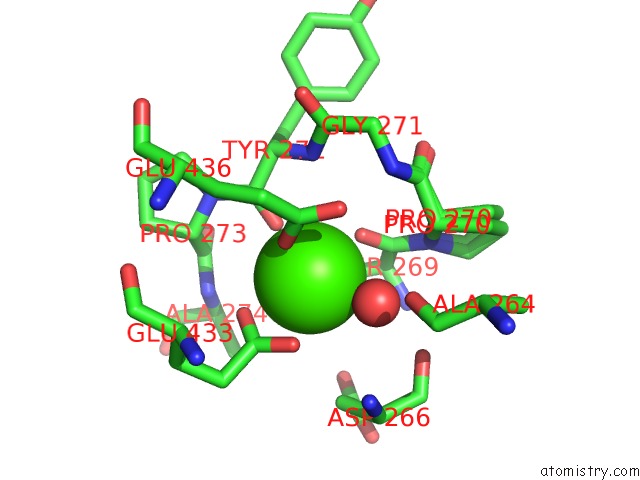
Mono view
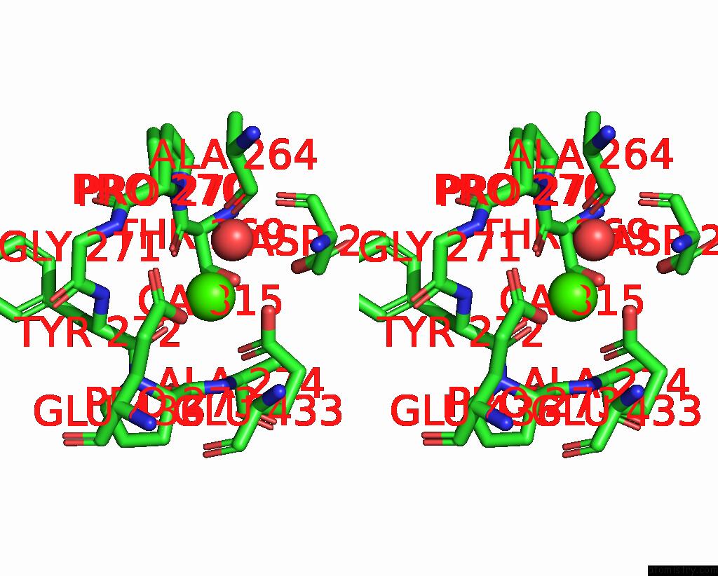
Stereo pair view
Mono view
Stereo pair view
A full contact list of Calcium with other atoms in the Ca binding
site number 1 of X-Ray Structure of Human Glutamate Carboxypeptidase II (Gcpii) in Complex with A Hydroxamate Inhibitor JHU241 within 5.0Å range:
|
Reference:
Z.Novakova,
K.Wozniak,
A.Jancarik,
R.Rais,
Y.Wu,
J.Pavlicek,
D.Ferraris,
B.Havlinova,
J.Ptacek,
J.Vavra,
N.Hin,
C.Rojas,
P.Majer,
B.S.Slusher,
T.Tsukamoto,
C.Barinka.
Unprecedented Binding Mode of Hydroxamate-Based Inhibitors of Glutamate Carboxypeptidase II: Structural Characterization and Biological Activity. J.Med.Chem. V. 59 4539 2016.
ISSN: ISSN 0022-2623
PubMed: 27074627
DOI: 10.1021/ACS.JMEDCHEM.5B01806
Page generated: Wed Jul 9 04:58:56 2025
ISSN: ISSN 0022-2623
PubMed: 27074627
DOI: 10.1021/ACS.JMEDCHEM.5B01806
Last articles
Fe in 2YXOFe in 2YRS
Fe in 2YXC
Fe in 2YNM
Fe in 2YVJ
Fe in 2YP1
Fe in 2YU2
Fe in 2YU1
Fe in 2YQB
Fe in 2YOO