Calcium »
PDB 5cy4-5dau »
5d62 »
Calcium in PDB 5d62: Moa-Z-Vad-Fmk Complex, Inverted Orientation
Protein crystallography data
The structure of Moa-Z-Vad-Fmk Complex, Inverted Orientation, PDB code: 5d62
was solved by
G.Cordara,
U.Krengel,
with X-Ray Crystallography technique. A brief refinement statistics is given in the table below:
| Resolution Low / High (Å) | 46.41 / 1.70 |
| Space group | P 63 2 2 |
| Cell size a, b, c (Å), α, β, γ (°) | 120.998, 120.998, 99.957, 90.00, 90.00, 120.00 |
| R / Rfree (%) | 16.9 / 19.8 |
Other elements in 5d62:
The structure of Moa-Z-Vad-Fmk Complex, Inverted Orientation also contains other interesting chemical elements:
| Chlorine | (Cl) | 2 atoms |
Calcium Binding Sites:
The binding sites of Calcium atom in the Moa-Z-Vad-Fmk Complex, Inverted Orientation
(pdb code 5d62). This binding sites where shown within
5.0 Angstroms radius around Calcium atom.
In total 2 binding sites of Calcium where determined in the Moa-Z-Vad-Fmk Complex, Inverted Orientation, PDB code: 5d62:
Jump to Calcium binding site number: 1; 2;
In total 2 binding sites of Calcium where determined in the Moa-Z-Vad-Fmk Complex, Inverted Orientation, PDB code: 5d62:
Jump to Calcium binding site number: 1; 2;
Calcium binding site 1 out of 2 in 5d62
Go back to
Calcium binding site 1 out
of 2 in the Moa-Z-Vad-Fmk Complex, Inverted Orientation
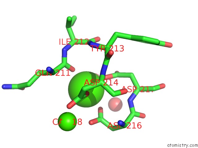
Mono view
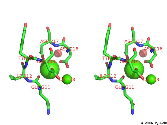
Stereo pair view
Mono view
Stereo pair view
A full contact list of Calcium with other atoms in the Ca binding
site number 1 of Moa-Z-Vad-Fmk Complex, Inverted Orientation within 5.0Å range:
|
Calcium binding site 2 out of 2 in 5d62
Go back to
Calcium binding site 2 out
of 2 in the Moa-Z-Vad-Fmk Complex, Inverted Orientation
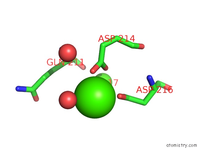
Mono view
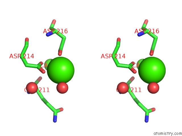
Stereo pair view
Mono view
Stereo pair view
A full contact list of Calcium with other atoms in the Ca binding
site number 2 of Moa-Z-Vad-Fmk Complex, Inverted Orientation within 5.0Å range:
|
Reference:
G.Cordara,
A.Van Eerde,
E.M.Grahn,
H.C.Winter,
I.J.Goldstein,
U.Krengel.
An Unusual Member of the Papain Superfamily: Mapping the Catalytic Cleft of the Marasmius Oreades Agglutinin (Moa) with A Caspase Inhibitor. Plos One V. 11 49407 2016.
ISSN: ESSN 1932-6203
PubMed: 26901797
DOI: 10.1371/JOURNAL.PONE.0149407
Page generated: Wed Jul 9 05:01:18 2025
ISSN: ESSN 1932-6203
PubMed: 26901797
DOI: 10.1371/JOURNAL.PONE.0149407
Last articles
Fe in 2YXOFe in 2YRS
Fe in 2YXC
Fe in 2YNM
Fe in 2YVJ
Fe in 2YP1
Fe in 2YU2
Fe in 2YU1
Fe in 2YQB
Fe in 2YOO