Calcium »
PDB 5dau-5dy1 »
5dcg »
Calcium in PDB 5dcg: Crystal Structure of Wt Apo Human Glutathione Transferase Pi
Enzymatic activity of Crystal Structure of Wt Apo Human Glutathione Transferase Pi
All present enzymatic activity of Crystal Structure of Wt Apo Human Glutathione Transferase Pi:
2.5.1.18;
2.5.1.18;
Protein crystallography data
The structure of Crystal Structure of Wt Apo Human Glutathione Transferase Pi, PDB code: 5dcg
was solved by
L.J.Parker,
M.W.Parker,
C.J.Morton,
with X-Ray Crystallography technique. A brief refinement statistics is given in the table below:
| Resolution Low / High (Å) | 38.33 / 2.01 |
| Space group | C 1 2 1 |
| Cell size a, b, c (Å), α, β, γ (°) | 77.499, 90.250, 68.920, 90.00, 98.49, 90.00 |
| R / Rfree (%) | 14.8 / 19 |
Calcium Binding Sites:
The binding sites of Calcium atom in the Crystal Structure of Wt Apo Human Glutathione Transferase Pi
(pdb code 5dcg). This binding sites where shown within
5.0 Angstroms radius around Calcium atom.
In total only one binding site of Calcium was determined in the Crystal Structure of Wt Apo Human Glutathione Transferase Pi, PDB code: 5dcg:
In total only one binding site of Calcium was determined in the Crystal Structure of Wt Apo Human Glutathione Transferase Pi, PDB code: 5dcg:
Calcium binding site 1 out of 1 in 5dcg
Go back to
Calcium binding site 1 out
of 1 in the Crystal Structure of Wt Apo Human Glutathione Transferase Pi
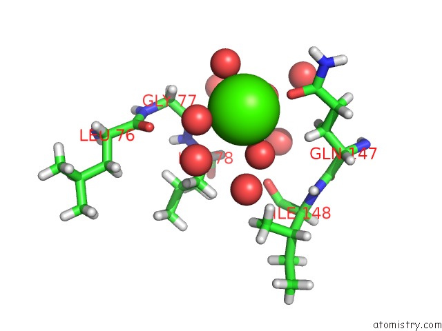
Mono view
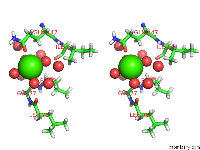
Stereo pair view
Mono view
Stereo pair view
A full contact list of Calcium with other atoms in the Ca binding
site number 1 of Crystal Structure of Wt Apo Human Glutathione Transferase Pi within 5.0Å range:
|
Reference:
L.J.Parker,
M.W.Parker,
C.J.Morton,
A.Bocedi,
D.B.Ascher,
J.B.Aitken,
H.H.Harris,
M.Lo Bello,
G.Ricci.
Visualisation of Organoarsenic Human Glutathione Transferase P1-1 Complexes: Metabolism of Arsenic-Based Therapeutics To Be Published.
Page generated: Sun Jul 14 17:59:32 2024
Last articles
Zn in 9JYWZn in 9IR4
Zn in 9IR3
Zn in 9GMX
Zn in 9GMW
Zn in 9JEJ
Zn in 9ERF
Zn in 9ERE
Zn in 9EGV
Zn in 9EGW