Calcium »
PDB 5day-5dzv »
5djk »
Calcium in PDB 5djk: Structure of M. Tuberculosis Cysq, A Pap Phosphatase with PO4 and 2CA Bound
Enzymatic activity of Structure of M. Tuberculosis Cysq, A Pap Phosphatase with PO4 and 2CA Bound
All present enzymatic activity of Structure of M. Tuberculosis Cysq, A Pap Phosphatase with PO4 and 2CA Bound:
3.1.3.11; 3.1.3.25; 3.1.3.7;
3.1.3.11; 3.1.3.25; 3.1.3.7;
Protein crystallography data
The structure of Structure of M. Tuberculosis Cysq, A Pap Phosphatase with PO4 and 2CA Bound, PDB code: 5djk
was solved by
A.J.Fisher,
A.I.Erickson,
with X-Ray Crystallography technique. A brief refinement statistics is given in the table below:
| Resolution Low / High (Å) | 31.53 / 1.80 |
| Space group | P 21 21 21 |
| Cell size a, b, c (Å), α, β, γ (°) | 40.257, 58.225, 101.415, 90.00, 90.00, 90.00 |
| R / Rfree (%) | 15.5 / 19 |
Other elements in 5djk:
The structure of Structure of M. Tuberculosis Cysq, A Pap Phosphatase with PO4 and 2CA Bound also contains other interesting chemical elements:
| Sodium | (Na) | 1 atom |
Calcium Binding Sites:
The binding sites of Calcium atom in the Structure of M. Tuberculosis Cysq, A Pap Phosphatase with PO4 and 2CA Bound
(pdb code 5djk). This binding sites where shown within
5.0 Angstroms radius around Calcium atom.
In total 2 binding sites of Calcium where determined in the Structure of M. Tuberculosis Cysq, A Pap Phosphatase with PO4 and 2CA Bound, PDB code: 5djk:
Jump to Calcium binding site number: 1; 2;
In total 2 binding sites of Calcium where determined in the Structure of M. Tuberculosis Cysq, A Pap Phosphatase with PO4 and 2CA Bound, PDB code: 5djk:
Jump to Calcium binding site number: 1; 2;
Calcium binding site 1 out of 2 in 5djk
Go back to
Calcium binding site 1 out
of 2 in the Structure of M. Tuberculosis Cysq, A Pap Phosphatase with PO4 and 2CA Bound

Mono view
Stereo pair view
Mono view
Stereo pair view
A full contact list of Calcium with other atoms in the Ca binding
site number 1 of Structure of M. Tuberculosis Cysq, A Pap Phosphatase with PO4 and 2CA Bound within 5.0Å range:
|
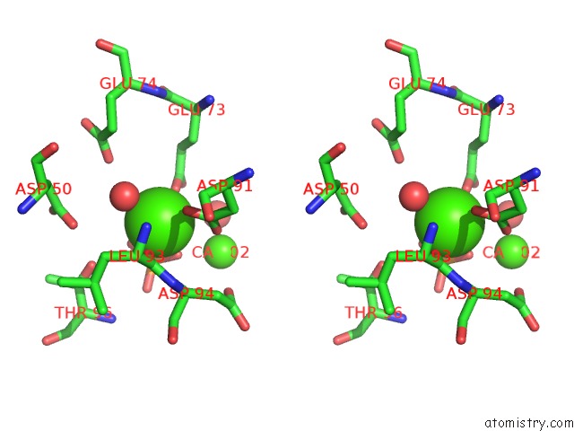
Calcium binding site 2 out of 2 in 5djk
Go back to
Calcium binding site 2 out
of 2 in the Structure of M. Tuberculosis Cysq, A Pap Phosphatase with PO4 and 2CA Bound

Mono view

Stereo pair view
Mono view
Stereo pair view
A full contact list of Calcium with other atoms in the Ca binding
site number 2 of Structure of M. Tuberculosis Cysq, A Pap Phosphatase with PO4 and 2CA Bound within 5.0Å range:
|
Reference:
A.I.Erickson,
R.D.Sarsam,
A.J.Fisher.
Crystal Structures of Mycobacterium Tuberculosis Cysq, with Substrate and Products Bound. Biochemistry V. 54 6830 2015.
ISSN: ISSN 0006-2960
PubMed: 26512869
DOI: 10.1021/ACS.BIOCHEM.5B01000
Page generated: Wed Jul 9 05:08:39 2025
ISSN: ISSN 0006-2960
PubMed: 26512869
DOI: 10.1021/ACS.BIOCHEM.5B01000
Last articles
Fe in 2YXOFe in 2YRS
Fe in 2YXC
Fe in 2YNM
Fe in 2YVJ
Fe in 2YP1
Fe in 2YU2
Fe in 2YU1
Fe in 2YQB
Fe in 2YOO