Calcium »
PDB 5day-5dzv »
5djq »
Calcium in PDB 5djq: The Structure of CBB3 Cytochrome Oxidase.
Enzymatic activity of The Structure of CBB3 Cytochrome Oxidase.
All present enzymatic activity of The Structure of CBB3 Cytochrome Oxidase.:
1.9.3.1;
1.9.3.1;
Protein crystallography data
The structure of The Structure of CBB3 Cytochrome Oxidase., PDB code: 5djq
was solved by
S.Buschmann,
E.Warkentin,
H.Xie,
M.Kohlstaedt,
J.D.Langer,
U.Ermler,
H.Michel,
with X-Ray Crystallography technique. A brief refinement statistics is given in the table below:
| Resolution Low / High (Å) | 14.98 / 3.20 |
| Space group | P 21 21 2 |
| Cell size a, b, c (Å), α, β, γ (°) | 136.475, 279.933, 175.192, 90.00, 90.00, 90.00 |
| R / Rfree (%) | 18.7 / 22.3 |
Other elements in 5djq:
The structure of The Structure of CBB3 Cytochrome Oxidase. also contains other interesting chemical elements:
| Iron | (Fe) | 24 atoms |
| Copper | (Cu) | 4 atoms |
Calcium Binding Sites:
The binding sites of Calcium atom in the The Structure of CBB3 Cytochrome Oxidase.
(pdb code 5djq). This binding sites where shown within
5.0 Angstroms radius around Calcium atom.
In total 8 binding sites of Calcium where determined in the The Structure of CBB3 Cytochrome Oxidase., PDB code: 5djq:
Jump to Calcium binding site number: 1; 2; 3; 4; 5; 6; 7; 8;
In total 8 binding sites of Calcium where determined in the The Structure of CBB3 Cytochrome Oxidase., PDB code: 5djq:
Jump to Calcium binding site number: 1; 2; 3; 4; 5; 6; 7; 8;
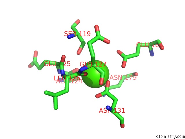
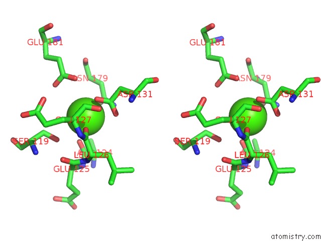
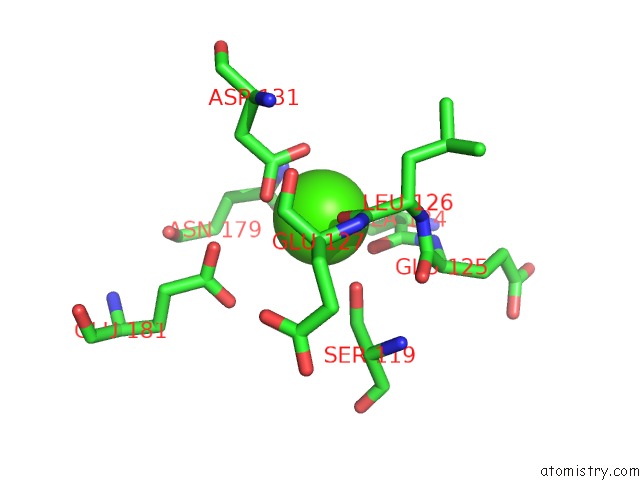
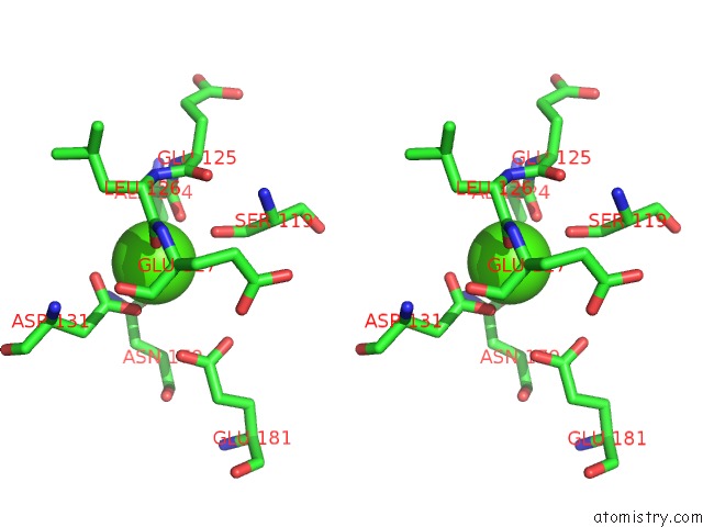
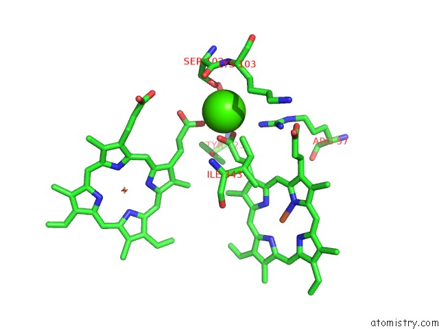
Calcium binding site 1 out of 8 in 5djq
Go back to
Calcium binding site 1 out
of 8 in the The Structure of CBB3 Cytochrome Oxidase.
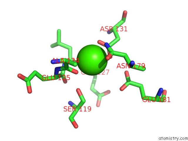
Mono view
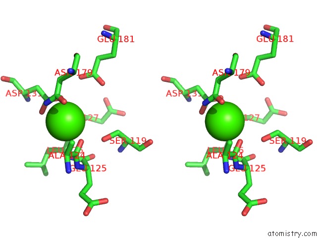
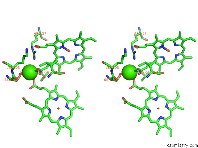
Stereo pair view
Mono view
Stereo pair view
A full contact list of Calcium with other atoms in the Ca binding
site number 1 of The Structure of CBB3 Cytochrome Oxidase. within 5.0Å range:
|
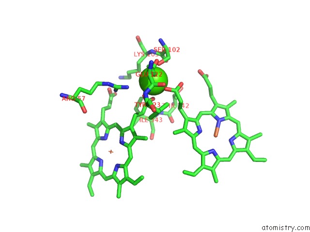
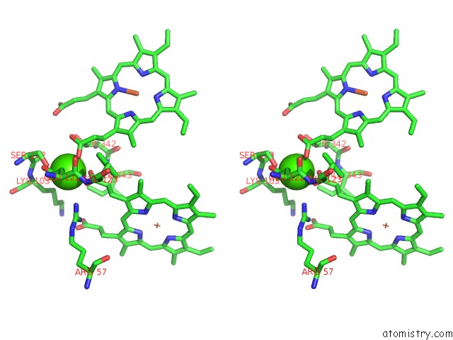
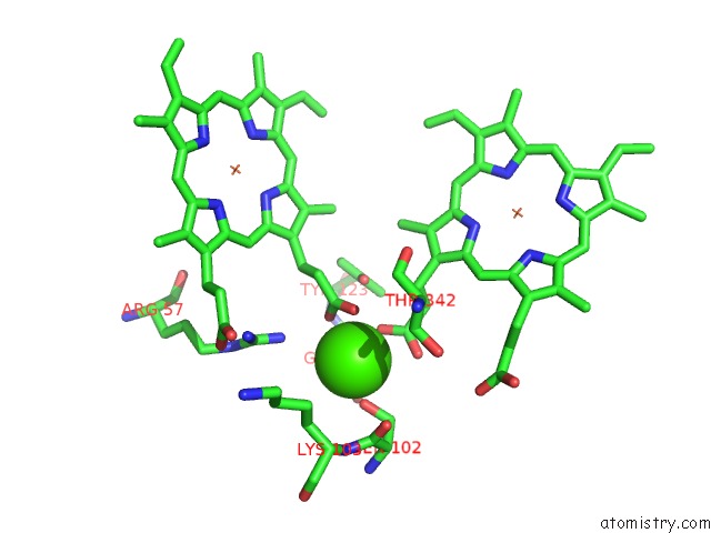
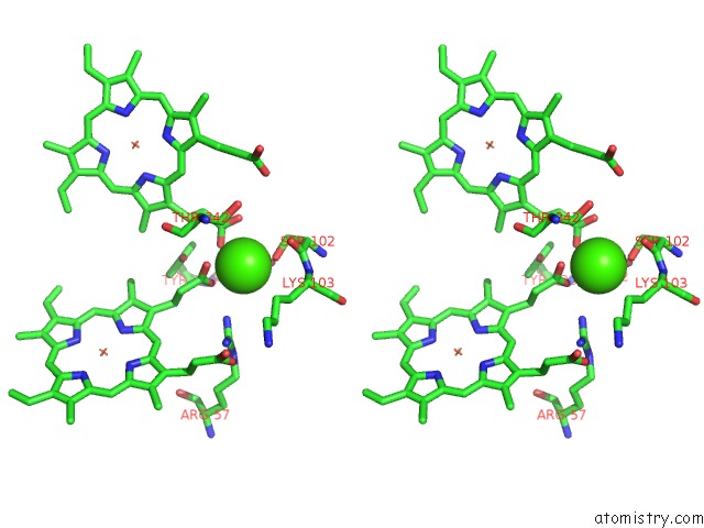
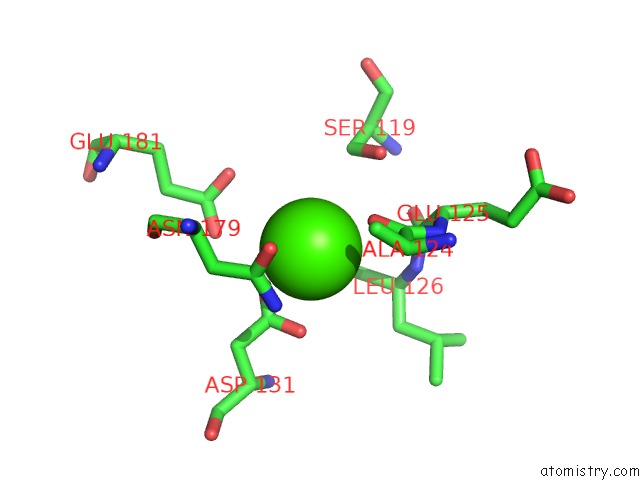
Calcium binding site 2 out of 8 in 5djq
Go back to
Calcium binding site 2 out
of 8 in the The Structure of CBB3 Cytochrome Oxidase.
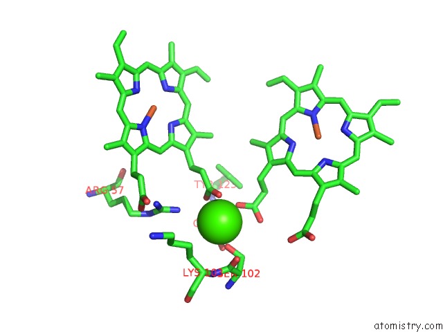
Mono view
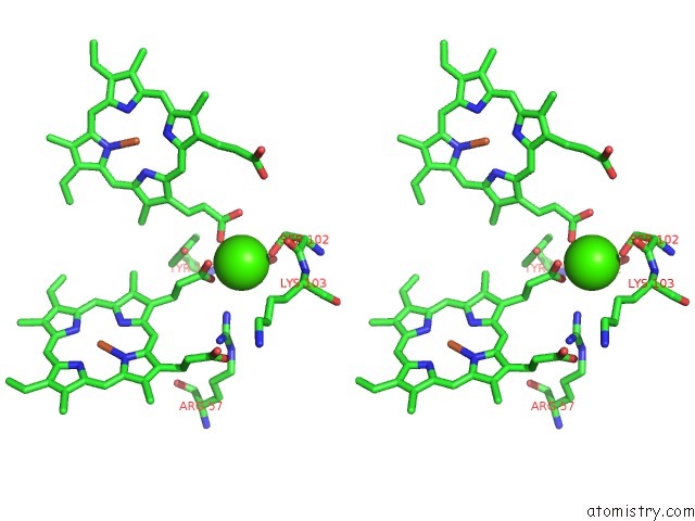
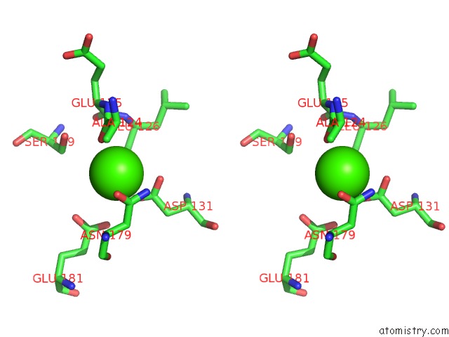
Stereo pair view
Mono view
Stereo pair view
A full contact list of Calcium with other atoms in the Ca binding
site number 2 of The Structure of CBB3 Cytochrome Oxidase. within 5.0Å range:
|
Calcium binding site 3 out of 8 in 5djq
Go back to
Calcium binding site 3 out
of 8 in the The Structure of CBB3 Cytochrome Oxidase.
Mono view
Stereo pair view
Mono view
Stereo pair view
A full contact list of Calcium with other atoms in the Ca binding
site number 3 of The Structure of CBB3 Cytochrome Oxidase. within 5.0Å range:
|
Calcium binding site 4 out of 8 in 5djq
Go back to
Calcium binding site 4 out
of 8 in the The Structure of CBB3 Cytochrome Oxidase.
Mono view
Stereo pair view
Mono view
Stereo pair view
A full contact list of Calcium with other atoms in the Ca binding
site number 4 of The Structure of CBB3 Cytochrome Oxidase. within 5.0Å range:
|
Calcium binding site 5 out of 8 in 5djq
Go back to
Calcium binding site 5 out
of 8 in the The Structure of CBB3 Cytochrome Oxidase.
Mono view
Stereo pair view
Mono view
Stereo pair view
A full contact list of Calcium with other atoms in the Ca binding
site number 5 of The Structure of CBB3 Cytochrome Oxidase. within 5.0Å range:
|
Calcium binding site 6 out of 8 in 5djq
Go back to
Calcium binding site 6 out
of 8 in the The Structure of CBB3 Cytochrome Oxidase.
Mono view
Stereo pair view
Mono view
Stereo pair view
A full contact list of Calcium with other atoms in the Ca binding
site number 6 of The Structure of CBB3 Cytochrome Oxidase. within 5.0Å range:
|
Calcium binding site 7 out of 8 in 5djq
Go back to
Calcium binding site 7 out
of 8 in the The Structure of CBB3 Cytochrome Oxidase.
Mono view
Stereo pair view
Mono view
Stereo pair view
A full contact list of Calcium with other atoms in the Ca binding
site number 7 of The Structure of CBB3 Cytochrome Oxidase. within 5.0Å range:
|
Calcium binding site 8 out of 8 in 5djq
Go back to
Calcium binding site 8 out
of 8 in the The Structure of CBB3 Cytochrome Oxidase.
Mono view
Stereo pair view
Mono view
Stereo pair view
A full contact list of Calcium with other atoms in the Ca binding
site number 8 of The Structure of CBB3 Cytochrome Oxidase. within 5.0Å range:
|
Reference:
S.Buschmann,
E.Warkentin,
H.Xie,
J.D.Langer,
U.Ermler,
H.Michel.
The Structure of CBB3 Cytochrome Oxidase Provides Insights Into Proton Pumping. Science V. 329 327 2010.
ISSN: ESSN 1095-9203
PubMed: 20576851
DOI: 10.1126/SCIENCE.1187303
Page generated: Sun Jul 14 18:01:29 2024
ISSN: ESSN 1095-9203
PubMed: 20576851
DOI: 10.1126/SCIENCE.1187303
Last articles
Zn in 9J0NZn in 9J0O
Zn in 9J0P
Zn in 9FJX
Zn in 9EKB
Zn in 9C0F
Zn in 9CAH
Zn in 9CH0
Zn in 9CH3
Zn in 9CH1