Calcium »
PDB 5day-5dzv »
5dl7 »
Calcium in PDB 5dl7: Crystal Structure of Acinetobacter Baumannii OCCAB3
Protein crystallography data
The structure of Crystal Structure of Acinetobacter Baumannii OCCAB3, PDB code: 5dl7
was solved by
M.Zahn,
A.Basle,
B.Van Den Berg,
with X-Ray Crystallography technique. A brief refinement statistics is given in the table below:
| Resolution Low / High (Å) | 70.14 / 1.75 |
| Space group | I 2 2 2 |
| Cell size a, b, c (Å), α, β, γ (°) | 81.563, 125.717, 137.459, 90.00, 90.00, 90.00 |
| R / Rfree (%) | 16.8 / 19.1 |
Calcium Binding Sites:
The binding sites of Calcium atom in the Crystal Structure of Acinetobacter Baumannii OCCAB3
(pdb code 5dl7). This binding sites where shown within
5.0 Angstroms radius around Calcium atom.
In total 4 binding sites of Calcium where determined in the Crystal Structure of Acinetobacter Baumannii OCCAB3, PDB code: 5dl7:
Jump to Calcium binding site number: 1; 2; 3; 4;
In total 4 binding sites of Calcium where determined in the Crystal Structure of Acinetobacter Baumannii OCCAB3, PDB code: 5dl7:
Jump to Calcium binding site number: 1; 2; 3; 4;
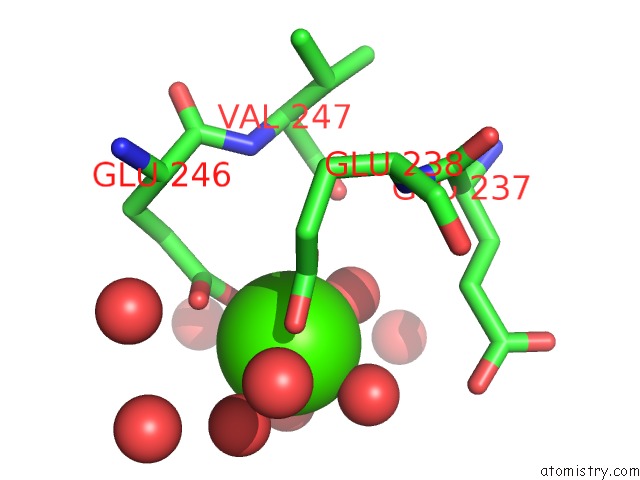
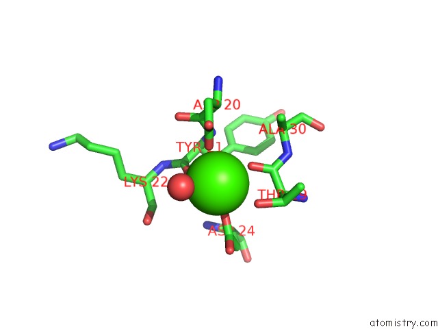
Calcium binding site 1 out of 4 in 5dl7
Go back to
Calcium binding site 1 out
of 4 in the Crystal Structure of Acinetobacter Baumannii OCCAB3
Mono view
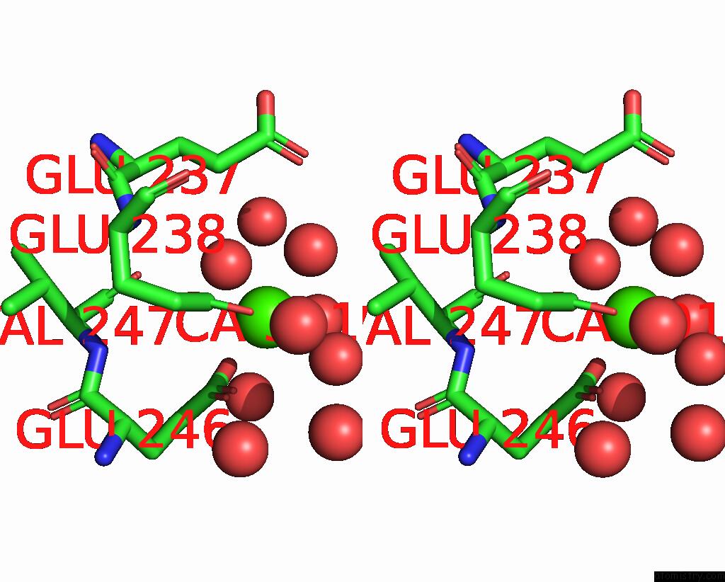
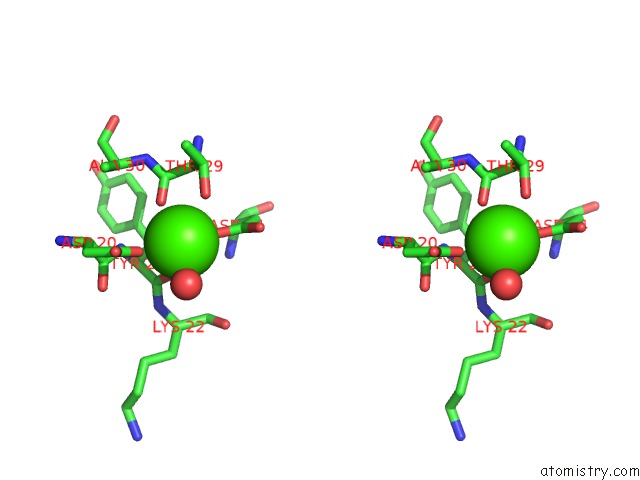
Stereo pair view
Mono view
Stereo pair view
A full contact list of Calcium with other atoms in the Ca binding
site number 1 of Crystal Structure of Acinetobacter Baumannii OCCAB3 within 5.0Å range:
|
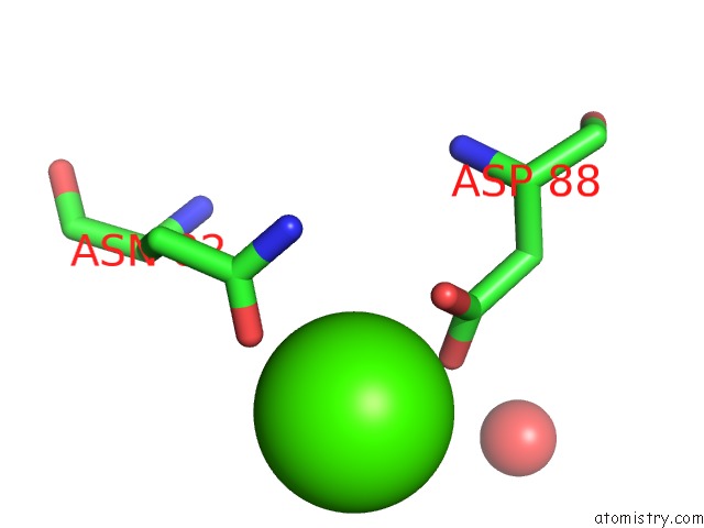
Calcium binding site 2 out of 4 in 5dl7
Go back to
Calcium binding site 2 out
of 4 in the Crystal Structure of Acinetobacter Baumannii OCCAB3
Mono view
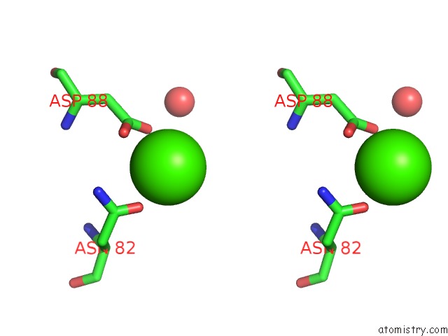
Stereo pair view
Mono view
Stereo pair view
A full contact list of Calcium with other atoms in the Ca binding
site number 2 of Crystal Structure of Acinetobacter Baumannii OCCAB3 within 5.0Å range:
|
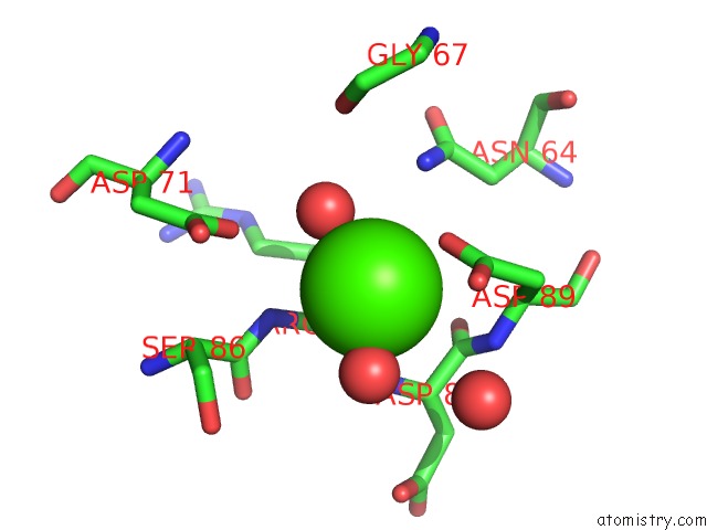
Calcium binding site 3 out of 4 in 5dl7
Go back to
Calcium binding site 3 out
of 4 in the Crystal Structure of Acinetobacter Baumannii OCCAB3
Mono view
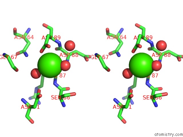
Stereo pair view
Mono view
Stereo pair view
A full contact list of Calcium with other atoms in the Ca binding
site number 3 of Crystal Structure of Acinetobacter Baumannii OCCAB3 within 5.0Å range:
|
Calcium binding site 4 out of 4 in 5dl7
Go back to
Calcium binding site 4 out
of 4 in the Crystal Structure of Acinetobacter Baumannii OCCAB3
Mono view
Stereo pair view
Mono view
Stereo pair view
A full contact list of Calcium with other atoms in the Ca binding
site number 4 of Crystal Structure of Acinetobacter Baumannii OCCAB3 within 5.0Å range:
|
Reference:
M.Zahn,
S.P.Bhamidimarri,
A.Basle,
M.Winterhalter,
B.Van Den Berg.
Structural Insights Into Outer Membrane Permeability of Acinetobacter Baumannii. Structure V. 24 221 2016.
ISSN: ISSN 0969-2126
PubMed: 26805524
DOI: 10.1016/J.STR.2015.12.009
Page generated: Sun Jul 14 18:02:58 2024
ISSN: ISSN 0969-2126
PubMed: 26805524
DOI: 10.1016/J.STR.2015.12.009
Last articles
Zn in 9J0NZn in 9J0O
Zn in 9J0P
Zn in 9FJX
Zn in 9EKB
Zn in 9C0F
Zn in 9CAH
Zn in 9CH0
Zn in 9CH3
Zn in 9CH1