Calcium »
PDB 5day-5dzv »
5doo »
Calcium in PDB 5doo: The Structure of PKMT2 From Rickettsia Typhi
Protein crystallography data
The structure of The Structure of PKMT2 From Rickettsia Typhi, PDB code: 5doo
was solved by
N.Noinaj,
A.Abeykoon,
Y.He,
D.C.Yang,
S.K.Buchanan,
with X-Ray Crystallography technique. A brief refinement statistics is given in the table below:
| Resolution Low / High (Å) | 48.45 / 3.13 |
| Space group | P 1 21 1 |
| Cell size a, b, c (Å), α, β, γ (°) | 83.719, 90.528, 106.201, 90.00, 114.17, 90.00 |
| R / Rfree (%) | 23.3 / 28 |
Calcium Binding Sites:
The binding sites of Calcium atom in the The Structure of PKMT2 From Rickettsia Typhi
(pdb code 5doo). This binding sites where shown within
5.0 Angstroms radius around Calcium atom.
In total 2 binding sites of Calcium where determined in the The Structure of PKMT2 From Rickettsia Typhi, PDB code: 5doo:
Jump to Calcium binding site number: 1; 2;
In total 2 binding sites of Calcium where determined in the The Structure of PKMT2 From Rickettsia Typhi, PDB code: 5doo:
Jump to Calcium binding site number: 1; 2;
Calcium binding site 1 out of 2 in 5doo
Go back to
Calcium binding site 1 out
of 2 in the The Structure of PKMT2 From Rickettsia Typhi
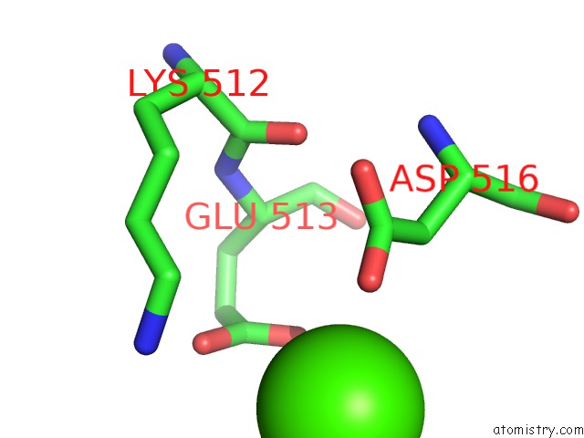
Mono view
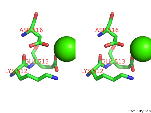
Stereo pair view
Mono view
Stereo pair view
A full contact list of Calcium with other atoms in the Ca binding
site number 1 of The Structure of PKMT2 From Rickettsia Typhi within 5.0Å range:
|
Calcium binding site 2 out of 2 in 5doo
Go back to
Calcium binding site 2 out
of 2 in the The Structure of PKMT2 From Rickettsia Typhi
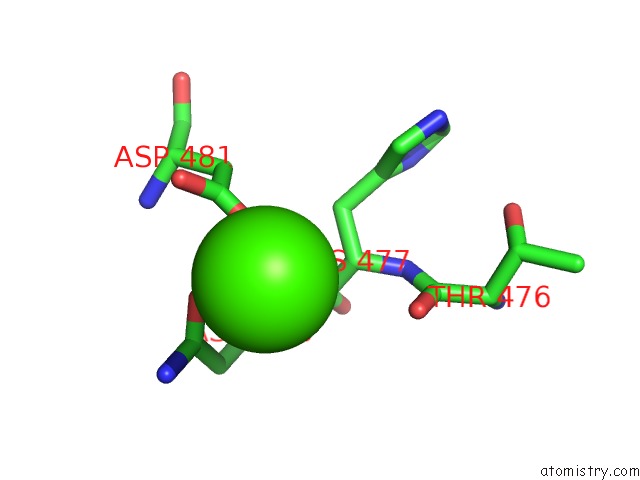
Mono view
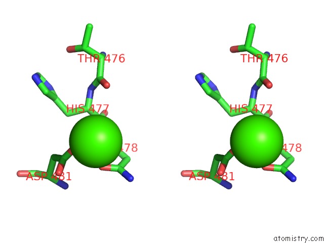
Stereo pair view
Mono view
Stereo pair view
A full contact list of Calcium with other atoms in the Ca binding
site number 2 of The Structure of PKMT2 From Rickettsia Typhi within 5.0Å range:
|
Reference:
A.H.Abeykoon,
N.Noinaj,
B.E.Choi,
L.Wise,
Y.He,
C.C.Chao,
G.Wang,
M.Gucek,
W.M.Ching,
P.B.Chock,
S.K.Buchanan,
D.C.Yang.
Structural Insights Into Substrate Recognition and Catalysis in Outer Membrane Protein B (Ompb) By Protein-Lysine Methyltransferases From Rickettsia. J.Biol.Chem. V. 291 19962 2016.
ISSN: ESSN 1083-351X
PubMed: 27474738
DOI: 10.1074/JBC.M116.723460
Page generated: Sun Jul 14 18:06:17 2024
ISSN: ESSN 1083-351X
PubMed: 27474738
DOI: 10.1074/JBC.M116.723460
Last articles
Zn in 9J0NZn in 9J0O
Zn in 9J0P
Zn in 9FJX
Zn in 9EKB
Zn in 9C0F
Zn in 9CAH
Zn in 9CH0
Zn in 9CH3
Zn in 9CH1