Calcium »
PDB 5dzw-5egt »
5e5o »
Calcium in PDB 5e5o: I-Smami Bound to Uncleaved Dna Target in the Presence of Calcium Ions
Protein crystallography data
The structure of I-Smami Bound to Uncleaved Dna Target in the Presence of Calcium Ions, PDB code: 5e5o
was solved by
B.W.Shen,
with X-Ray Crystallography technique. A brief refinement statistics is given in the table below:
| Resolution Low / High (Å) | 27.00 / 2.36 |
| Space group | P 21 21 21 |
| Cell size a, b, c (Å), α, β, γ (°) | 61.327, 67.993, 97.743, 90.00, 90.00, 90.00 |
| R / Rfree (%) | 18.8 / 27.4 |
Calcium Binding Sites:
The binding sites of Calcium atom in the I-Smami Bound to Uncleaved Dna Target in the Presence of Calcium Ions
(pdb code 5e5o). This binding sites where shown within
5.0 Angstroms radius around Calcium atom.
In total 3 binding sites of Calcium where determined in the I-Smami Bound to Uncleaved Dna Target in the Presence of Calcium Ions, PDB code: 5e5o:
Jump to Calcium binding site number: 1; 2; 3;
In total 3 binding sites of Calcium where determined in the I-Smami Bound to Uncleaved Dna Target in the Presence of Calcium Ions, PDB code: 5e5o:
Jump to Calcium binding site number: 1; 2; 3;
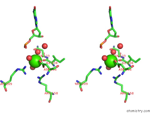
Calcium binding site 1 out of 3 in 5e5o
Go back to
Calcium binding site 1 out
of 3 in the I-Smami Bound to Uncleaved Dna Target in the Presence of Calcium Ions

Mono view
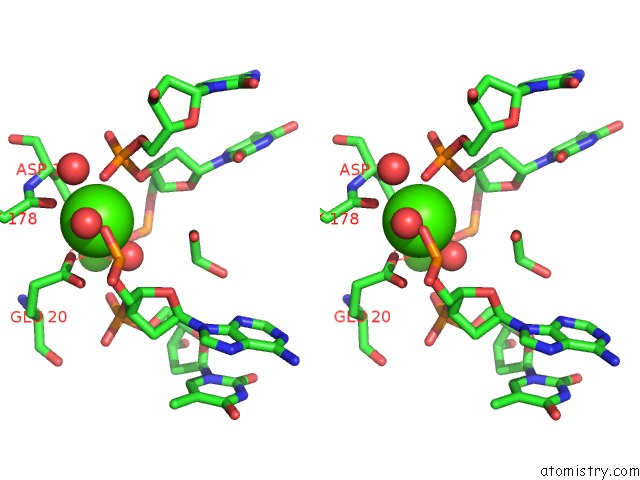
Stereo pair view
Mono view
Stereo pair view
A full contact list of Calcium with other atoms in the Ca binding
site number 1 of I-Smami Bound to Uncleaved Dna Target in the Presence of Calcium Ions within 5.0Å range:
|
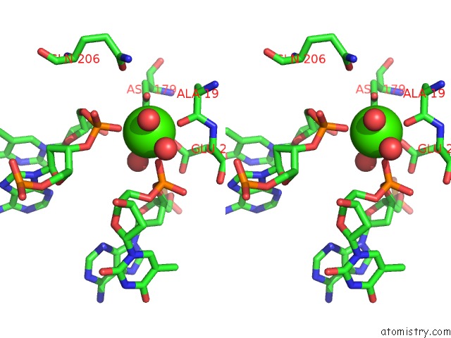
Calcium binding site 2 out of 3 in 5e5o
Go back to
Calcium binding site 2 out
of 3 in the I-Smami Bound to Uncleaved Dna Target in the Presence of Calcium Ions
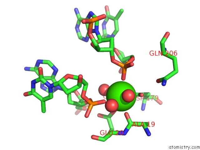
Mono view
Stereo pair view
Mono view
Stereo pair view
A full contact list of Calcium with other atoms in the Ca binding
site number 2 of I-Smami Bound to Uncleaved Dna Target in the Presence of Calcium Ions within 5.0Å range:
|
Calcium binding site 3 out of 3 in 5e5o
Go back to
Calcium binding site 3 out
of 3 in the I-Smami Bound to Uncleaved Dna Target in the Presence of Calcium Ions
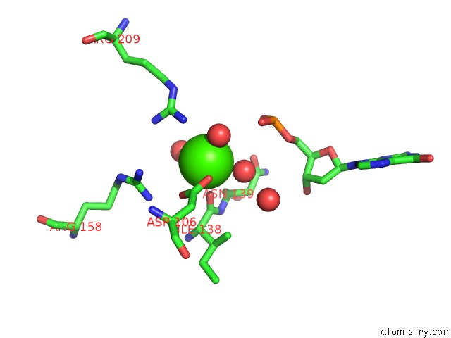
Mono view
Stereo pair view
Mono view
Stereo pair view
A full contact list of Calcium with other atoms in the Ca binding
site number 3 of I-Smami Bound to Uncleaved Dna Target in the Presence of Calcium Ions within 5.0Å range:
|
Reference:
B.W.Shen,
A.Lambert,
B.C.Walker,
B.L.Stoddard,
B.K.Kaiser.
The Structural Basis of Asymmetry in Dna Binding and Cleavage As Exhibited By the I-Smami Laglidadg Meganuclease. J.Mol.Biol. V. 428 206 2016.
ISSN: ESSN 1089-8638
PubMed: 26705195
DOI: 10.1016/J.JMB.2015.12.005
Page generated: Wed Jul 9 05:26:04 2025
ISSN: ESSN 1089-8638
PubMed: 26705195
DOI: 10.1016/J.JMB.2015.12.005
Last articles
Cl in 5G54Cl in 5G4A
Cl in 5G4Q
Cl in 5G47
Cl in 5G42
Cl in 5G3S
Cl in 5G2P
Cl in 5G2T
Cl in 5G36
Cl in 5G2D