Calcium »
PDB 5dzw-5egt »
5e6u »
Calcium in PDB 5e6u: Structures of Leukocyte Integrin ALB2: the Ai Domain, the Headpiece, and the Pocket For the Internal Ligand
Protein crystallography data
The structure of Structures of Leukocyte Integrin ALB2: the Ai Domain, the Headpiece, and the Pocket For the Internal Ligand, PDB code: 5e6u
was solved by
M.Sen,
T.A.Springer,
with X-Ray Crystallography technique. A brief refinement statistics is given in the table below:
| Resolution Low / High (Å) | 46.44 / 2.50 |
| Space group | P 61 |
| Cell size a, b, c (Å), α, β, γ (°) | 154.980, 154.980, 115.450, 90.00, 90.00, 120.00 |
| R / Rfree (%) | 17.9 / 22.6 |
Other elements in 5e6u:
The structure of Structures of Leukocyte Integrin ALB2: the Ai Domain, the Headpiece, and the Pocket For the Internal Ligand also contains other interesting chemical elements:
| Magnesium | (Mg) | 2 atoms |
| Chlorine | (Cl) | 1 atom |
Calcium Binding Sites:
The binding sites of Calcium atom in the Structures of Leukocyte Integrin ALB2: the Ai Domain, the Headpiece, and the Pocket For the Internal Ligand
(pdb code 5e6u). This binding sites where shown within
5.0 Angstroms radius around Calcium atom.
In total 4 binding sites of Calcium where determined in the Structures of Leukocyte Integrin ALB2: the Ai Domain, the Headpiece, and the Pocket For the Internal Ligand, PDB code: 5e6u:
Jump to Calcium binding site number: 1; 2; 3; 4;
In total 4 binding sites of Calcium where determined in the Structures of Leukocyte Integrin ALB2: the Ai Domain, the Headpiece, and the Pocket For the Internal Ligand, PDB code: 5e6u:
Jump to Calcium binding site number: 1; 2; 3; 4;
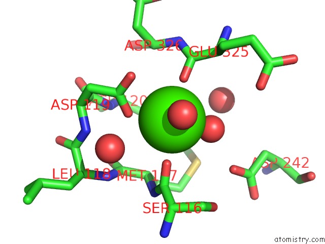
Calcium binding site 1 out of 4 in 5e6u
Go back to
Calcium binding site 1 out
of 4 in the Structures of Leukocyte Integrin ALB2: the Ai Domain, the Headpiece, and the Pocket For the Internal Ligand
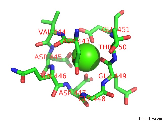
Mono view
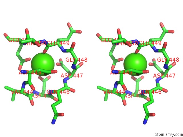
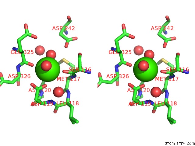
Stereo pair view
Mono view
Stereo pair view
A full contact list of Calcium with other atoms in the Ca binding
site number 1 of Structures of Leukocyte Integrin ALB2: the Ai Domain, the Headpiece, and the Pocket For the Internal Ligand within 5.0Å range:
|
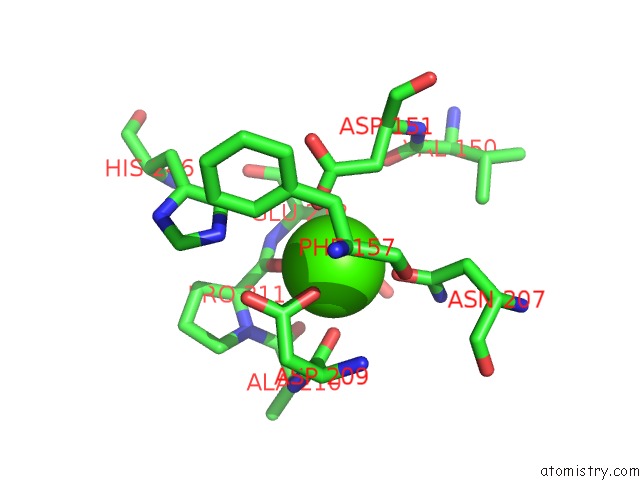
Calcium binding site 2 out of 4 in 5e6u
Go back to
Calcium binding site 2 out
of 4 in the Structures of Leukocyte Integrin ALB2: the Ai Domain, the Headpiece, and the Pocket For the Internal Ligand
Mono view
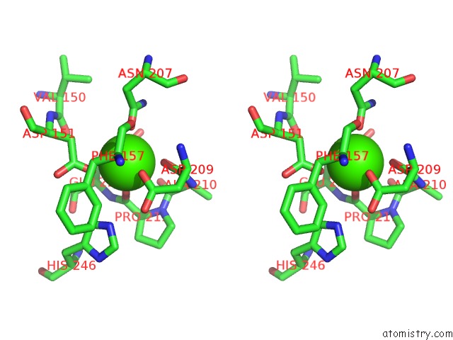
Stereo pair view
Mono view
Stereo pair view
A full contact list of Calcium with other atoms in the Ca binding
site number 2 of Structures of Leukocyte Integrin ALB2: the Ai Domain, the Headpiece, and the Pocket For the Internal Ligand within 5.0Å range:
|
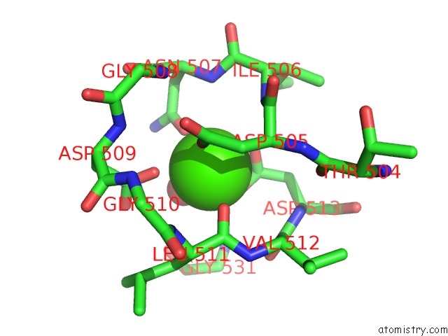
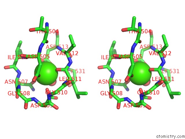
Calcium binding site 3 out of 4 in 5e6u
Go back to
Calcium binding site 3 out
of 4 in the Structures of Leukocyte Integrin ALB2: the Ai Domain, the Headpiece, and the Pocket For the Internal Ligand
Mono view
Stereo pair view
Mono view
Stereo pair view
A full contact list of Calcium with other atoms in the Ca binding
site number 3 of Structures of Leukocyte Integrin ALB2: the Ai Domain, the Headpiece, and the Pocket For the Internal Ligand within 5.0Å range:
|
Calcium binding site 4 out of 4 in 5e6u
Go back to
Calcium binding site 4 out
of 4 in the Structures of Leukocyte Integrin ALB2: the Ai Domain, the Headpiece, and the Pocket For the Internal Ligand
Mono view
Stereo pair view
Mono view
Stereo pair view
A full contact list of Calcium with other atoms in the Ca binding
site number 4 of Structures of Leukocyte Integrin ALB2: the Ai Domain, the Headpiece, and the Pocket For the Internal Ligand within 5.0Å range:
|
Reference:
M.Sen,
T.A.Springer.
Leukocyte Integrin Alpha L Beta 2 Headpiece Structures: the Alpha I Domain, the Pocket For the Internal Ligand, and Concerted Movements of Its Loops. Proc.Natl.Acad.Sci.Usa V. 113 2940 2016.
ISSN: ESSN 1091-6490
PubMed: 26936951
DOI: 10.1073/PNAS.1601379113
Page generated: Wed Jul 9 05:27:07 2025
ISSN: ESSN 1091-6490
PubMed: 26936951
DOI: 10.1073/PNAS.1601379113
Last articles
Fe in 2YXOFe in 2YRS
Fe in 2YXC
Fe in 2YNM
Fe in 2YVJ
Fe in 2YP1
Fe in 2YU2
Fe in 2YU1
Fe in 2YQB
Fe in 2YOO