Calcium »
PDB 5dzw-5egt »
5e7t »
Calcium in PDB 5e7t: Structure of the Tripod (Bppuct-A-L) From the Baseplate of Bacteriophage TUC2009
Protein crystallography data
The structure of Structure of the Tripod (Bppuct-A-L) From the Baseplate of Bacteriophage TUC2009, PDB code: 5e7t
was solved by
P.Legrand,
B.Collins,
S.Blangy,
J.Murphy,
S.Spinelli,
C.Gutierrez,
N.Richet,
C.Kellenberger,
A.Desmyter,
J.Mahony,
D.Van Sinderen,
C.Cambillau,
with X-Ray Crystallography technique. A brief refinement statistics is given in the table below:
| Resolution Low / High (Å) | 34.85 / 2.90 |
| Space group | P 21 3 |
| Cell size a, b, c (Å), α, β, γ (°) | 211.960, 211.960, 211.960, 90.00, 90.00, 90.00 |
| R / Rfree (%) | 21.4 / 23.7 |
Calcium Binding Sites:
The binding sites of Calcium atom in the Structure of the Tripod (Bppuct-A-L) From the Baseplate of Bacteriophage TUC2009
(pdb code 5e7t). This binding sites where shown within
5.0 Angstroms radius around Calcium atom.
In total only one binding site of Calcium was determined in the Structure of the Tripod (Bppuct-A-L) From the Baseplate of Bacteriophage TUC2009, PDB code: 5e7t:
In total only one binding site of Calcium was determined in the Structure of the Tripod (Bppuct-A-L) From the Baseplate of Bacteriophage TUC2009, PDB code: 5e7t:
Calcium binding site 1 out of 1 in 5e7t
Go back to
Calcium binding site 1 out
of 1 in the Structure of the Tripod (Bppuct-A-L) From the Baseplate of Bacteriophage TUC2009
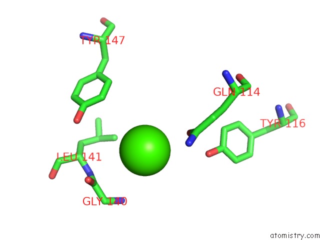
Mono view
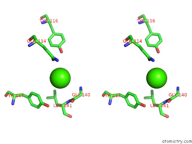
Stereo pair view
Mono view
Stereo pair view
A full contact list of Calcium with other atoms in the Ca binding
site number 1 of Structure of the Tripod (Bppuct-A-L) From the Baseplate of Bacteriophage TUC2009 within 5.0Å range:
|
Reference:
P.Legrand,
B.Collins,
S.Blangy,
J.Murphy,
S.Spinelli,
C.Gutierrez,
N.Richet,
C.Kellenberger,
A.Desmyter,
J.Mahony,
D.Van Sinderen,
C.Cambillau.
The Atomic Structure of the Phage TUC2009 Baseplate Tripod Suggests That Host Recognition Involves Two Different Carbohydrate Binding Modules. Mbio V. 7 01781 2016.
ISSN: ESSN 2150-7511
PubMed: 26814179
DOI: 10.1128/MBIO.01781-15
Page generated: Sun Jul 14 18:26:01 2024
ISSN: ESSN 2150-7511
PubMed: 26814179
DOI: 10.1128/MBIO.01781-15
Last articles
Zn in 9MJ5Zn in 9HNW
Zn in 9G0L
Zn in 9FNE
Zn in 9DZN
Zn in 9E0I
Zn in 9D32
Zn in 9DAK
Zn in 8ZXC
Zn in 8ZUF