Calcium »
PDB 5dzw-5egt »
5ebc »
Calcium in PDB 5ebc: Crystal Structure of ECCB1 of Mycobacterium Tuberculosis in Spacegroup P21 (State III)
Protein crystallography data
The structure of Crystal Structure of ECCB1 of Mycobacterium Tuberculosis in Spacegroup P21 (State III), PDB code: 5ebc
was solved by
X.L.Zhang,
C.Qi,
X.Q.Xie,
D.F.Li,
L.J.Bi,
with X-Ray Crystallography technique. A brief refinement statistics is given in the table below:
| Resolution Low / High (Å) | 30.28 / 3.00 |
| Space group | P 1 21 1 |
| Cell size a, b, c (Å), α, β, γ (°) | 31.610, 120.210, 61.260, 90.00, 102.92, 90.00 |
| R / Rfree (%) | 23.2 / 28.6 |
Calcium Binding Sites:
The binding sites of Calcium atom in the Crystal Structure of ECCB1 of Mycobacterium Tuberculosis in Spacegroup P21 (State III)
(pdb code 5ebc). This binding sites where shown within
5.0 Angstroms radius around Calcium atom.
In total only one binding site of Calcium was determined in the Crystal Structure of ECCB1 of Mycobacterium Tuberculosis in Spacegroup P21 (State III), PDB code: 5ebc:
In total only one binding site of Calcium was determined in the Crystal Structure of ECCB1 of Mycobacterium Tuberculosis in Spacegroup P21 (State III), PDB code: 5ebc:
Calcium binding site 1 out of 1 in 5ebc
Go back to
Calcium binding site 1 out
of 1 in the Crystal Structure of ECCB1 of Mycobacterium Tuberculosis in Spacegroup P21 (State III)
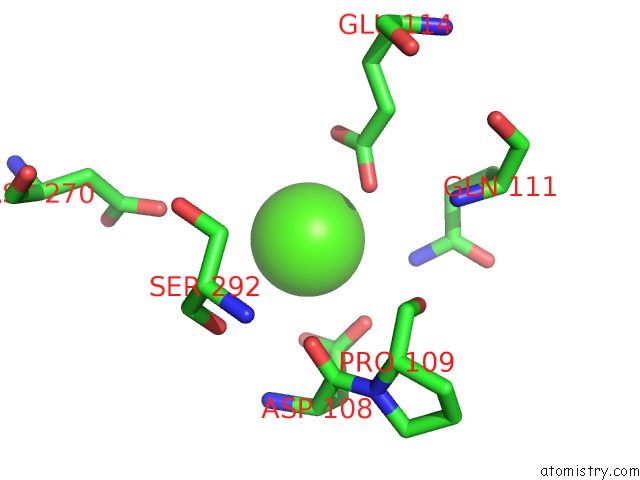
Mono view
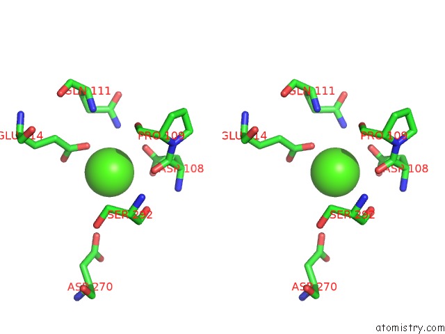
Stereo pair view
Mono view
Stereo pair view
A full contact list of Calcium with other atoms in the Ca binding
site number 1 of Crystal Structure of ECCB1 of Mycobacterium Tuberculosis in Spacegroup P21 (State III) within 5.0Å range:
|
Reference:
X.Q.Xie,
X.L.Zhang,
C.Qi,
D.F.Li,
J.Fleming,
D.C.Wang,
L.J.Bi.
Crystallographic Observation of the Movement of the Membrane-Distal Domain of the T7SS Core Component ECCB1 From Mycobacterium Tuberculosis. Acta Crystallogr.,Sect.F V. 72 139 2016.
ISSN: ESSN 2053-230X
PubMed: 26841765
DOI: 10.1107/S2053230X16000212
Page generated: Wed Jul 9 05:31:32 2025
ISSN: ESSN 2053-230X
PubMed: 26841765
DOI: 10.1107/S2053230X16000212
Last articles
Cl in 8B6PCl in 8B6R
Cl in 8B6Q
Cl in 8B6I
Cl in 8B6O
Cl in 8B5X
Cl in 8B5P
Cl in 8B2Y
Cl in 8B4X
Cl in 8B4W