Calcium »
PDB 5dzw-5egt »
5ed4 »
Calcium in PDB 5ed4: Structure of A Phop-Dna Complex
Enzymatic activity of Structure of A Phop-Dna Complex
All present enzymatic activity of Structure of A Phop-Dna Complex:
3.1.3.1;
3.1.3.1;
Protein crystallography data
The structure of Structure of A Phop-Dna Complex, PDB code: 5ed4
was solved by
S.Wang,
with X-Ray Crystallography technique. A brief refinement statistics is given in the table below:
| Resolution Low / High (Å) | 29.60 / 2.40 |
| Space group | P 21 21 21 |
| Cell size a, b, c (Å), α, β, γ (°) | 92.240, 98.176, 167.888, 90.00, 90.00, 90.00 |
| R / Rfree (%) | 18.3 / 22.7 |
Other elements in 5ed4:
The structure of Structure of A Phop-Dna Complex also contains other interesting chemical elements:
| Arsenic | (As) | 1 atom |
Calcium Binding Sites:
The binding sites of Calcium atom in the Structure of A Phop-Dna Complex
(pdb code 5ed4). This binding sites where shown within
5.0 Angstroms radius around Calcium atom.
In total 6 binding sites of Calcium where determined in the Structure of A Phop-Dna Complex, PDB code: 5ed4:
Jump to Calcium binding site number: 1; 2; 3; 4; 5; 6;
In total 6 binding sites of Calcium where determined in the Structure of A Phop-Dna Complex, PDB code: 5ed4:
Jump to Calcium binding site number: 1; 2; 3; 4; 5; 6;
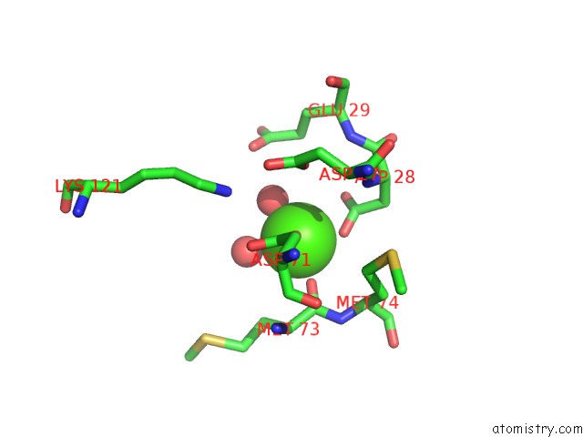
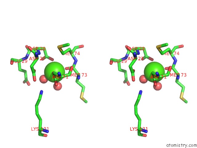
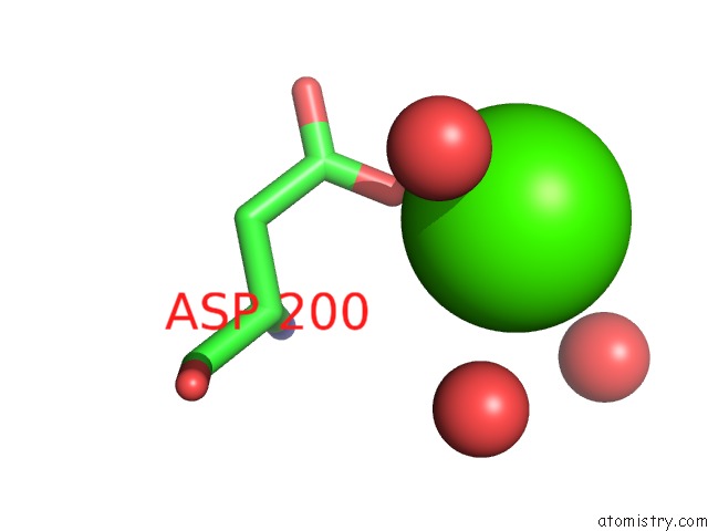
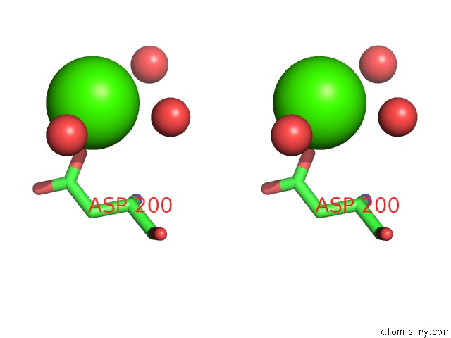
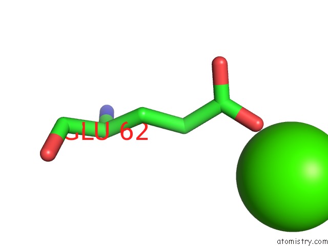
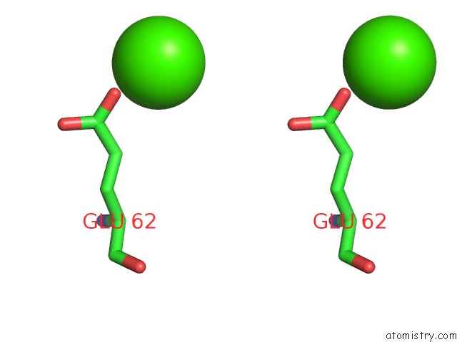
Calcium binding site 1 out of 6 in 5ed4
Go back to
Calcium binding site 1 out
of 6 in the Structure of A Phop-Dna Complex
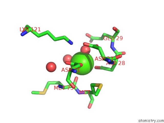
Mono view
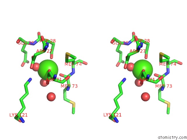
Stereo pair view
Mono view
Stereo pair view
A full contact list of Calcium with other atoms in the Ca binding
site number 1 of Structure of A Phop-Dna Complex within 5.0Å range:
|
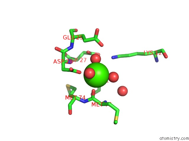
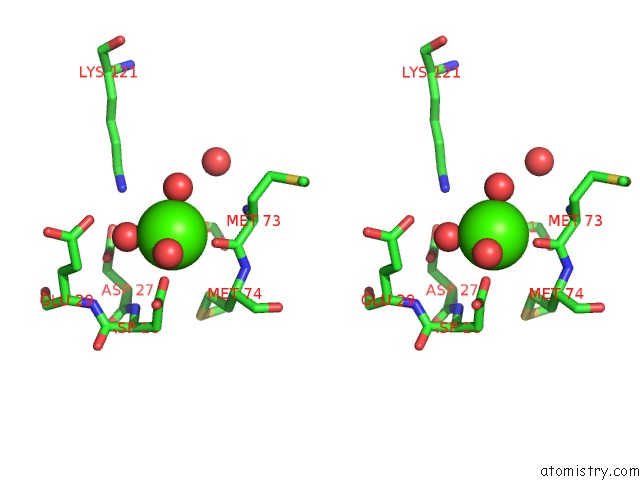
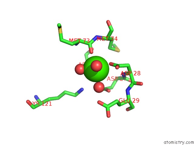
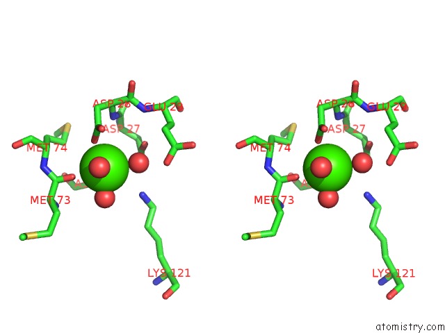
Calcium binding site 2 out of 6 in 5ed4
Go back to
Calcium binding site 2 out
of 6 in the Structure of A Phop-Dna Complex
Mono view
Stereo pair view
Mono view
Stereo pair view
A full contact list of Calcium with other atoms in the Ca binding
site number 2 of Structure of A Phop-Dna Complex within 5.0Å range:
|
Calcium binding site 3 out of 6 in 5ed4
Go back to
Calcium binding site 3 out
of 6 in the Structure of A Phop-Dna Complex
Mono view
Stereo pair view
Mono view
Stereo pair view
A full contact list of Calcium with other atoms in the Ca binding
site number 3 of Structure of A Phop-Dna Complex within 5.0Å range:
|
Calcium binding site 4 out of 6 in 5ed4
Go back to
Calcium binding site 4 out
of 6 in the Structure of A Phop-Dna Complex
Mono view
Stereo pair view
Mono view
Stereo pair view
A full contact list of Calcium with other atoms in the Ca binding
site number 4 of Structure of A Phop-Dna Complex within 5.0Å range:
|
Calcium binding site 5 out of 6 in 5ed4
Go back to
Calcium binding site 5 out
of 6 in the Structure of A Phop-Dna Complex
Mono view
Stereo pair view
Mono view
Stereo pair view
A full contact list of Calcium with other atoms in the Ca binding
site number 5 of Structure of A Phop-Dna Complex within 5.0Å range:
|
Calcium binding site 6 out of 6 in 5ed4
Go back to
Calcium binding site 6 out
of 6 in the Structure of A Phop-Dna Complex
Mono view
Stereo pair view
Mono view
Stereo pair view
A full contact list of Calcium with other atoms in the Ca binding
site number 6 of Structure of A Phop-Dna Complex within 5.0Å range:
|
Reference:
X.He,
L.Wang,
S.Wang.
Structural Basis of Dna Sequence Recognition By the Response Regulator Phop in Mycobacterium Tuberculosis. Sci Rep V. 6 24442 2016.
ISSN: ESSN 2045-2322
PubMed: 27079268
DOI: 10.1038/SREP24442
Page generated: Sun Jul 14 18:28:55 2024
ISSN: ESSN 2045-2322
PubMed: 27079268
DOI: 10.1038/SREP24442
Last articles
Zn in 9MJ5Zn in 9HNW
Zn in 9G0L
Zn in 9FNE
Zn in 9DZN
Zn in 9E0I
Zn in 9D32
Zn in 9DAK
Zn in 8ZXC
Zn in 8ZUF