Calcium »
PDB 5ekk-5eyh »
5ex2 »
Calcium in PDB 5ex2: Crystal Structure of Cyclophilin AQUACYP293 From Hirschia Baltica
Enzymatic activity of Crystal Structure of Cyclophilin AQUACYP293 From Hirschia Baltica
All present enzymatic activity of Crystal Structure of Cyclophilin AQUACYP293 From Hirschia Baltica:
5.2.1.8;
5.2.1.8;
Protein crystallography data
The structure of Crystal Structure of Cyclophilin AQUACYP293 From Hirschia Baltica, PDB code: 5ex2
was solved by
R.P.Jakob,
T.Maier,
with X-Ray Crystallography technique. A brief refinement statistics is given in the table below:
| Resolution Low / High (Å) | 47.87 / 1.29 |
| Space group | P 1 21 1 |
| Cell size a, b, c (Å), α, β, γ (°) | 47.940, 72.730, 73.930, 90.00, 93.00, 90.00 |
| R / Rfree (%) | 16.9 / 19.5 |
Other elements in 5ex2:
The structure of Crystal Structure of Cyclophilin AQUACYP293 From Hirschia Baltica also contains other interesting chemical elements:
| Magnesium | (Mg) | 2 atoms |
| Chlorine | (Cl) | 1 atom |
Calcium Binding Sites:
The binding sites of Calcium atom in the Crystal Structure of Cyclophilin AQUACYP293 From Hirschia Baltica
(pdb code 5ex2). This binding sites where shown within
5.0 Angstroms radius around Calcium atom.
In total only one binding site of Calcium was determined in the Crystal Structure of Cyclophilin AQUACYP293 From Hirschia Baltica, PDB code: 5ex2:
In total only one binding site of Calcium was determined in the Crystal Structure of Cyclophilin AQUACYP293 From Hirschia Baltica, PDB code: 5ex2:
Calcium binding site 1 out of 1 in 5ex2
Go back to
Calcium binding site 1 out
of 1 in the Crystal Structure of Cyclophilin AQUACYP293 From Hirschia Baltica
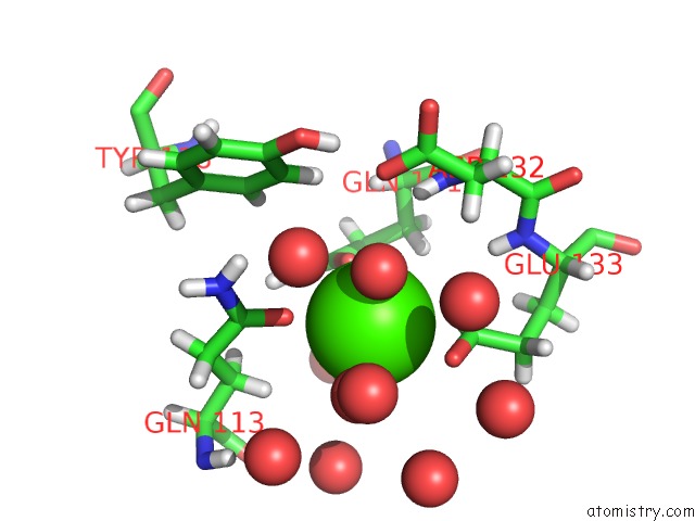
Mono view
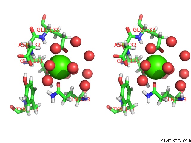
Stereo pair view
Mono view
Stereo pair view
A full contact list of Calcium with other atoms in the Ca binding
site number 1 of Crystal Structure of Cyclophilin AQUACYP293 From Hirschia Baltica within 5.0Å range:
|
Reference:
R.P.Jakob,
P.A.Schmidpeter,
J.R.Koch,
F.X.Schmid,
T.Maier.
Structural and Functional Characterization of A Novel Family of Cyclophilins, the Aquacyps. Plos One V. 11 57070 2016.
ISSN: ESSN 1932-6203
PubMed: 27276069
DOI: 10.1371/JOURNAL.PONE.0157070
Page generated: Sun Jul 14 18:54:31 2024
ISSN: ESSN 1932-6203
PubMed: 27276069
DOI: 10.1371/JOURNAL.PONE.0157070
Last articles
Zn in 9J0NZn in 9J0O
Zn in 9J0P
Zn in 9FJX
Zn in 9EKB
Zn in 9C0F
Zn in 9CAH
Zn in 9CH0
Zn in 9CH3
Zn in 9CH1