Calcium »
PDB 5eym-5fk0 »
5f6m »
Calcium in PDB 5f6m: Isotropic Trypsin Model For Comparison of Diffuse Scattering
Enzymatic activity of Isotropic Trypsin Model For Comparison of Diffuse Scattering
All present enzymatic activity of Isotropic Trypsin Model For Comparison of Diffuse Scattering:
3.4.21.4;
3.4.21.4;
Protein crystallography data
The structure of Isotropic Trypsin Model For Comparison of Diffuse Scattering, PDB code: 5f6m
was solved by
A.H.Van Benschoten,
M.E.Wall,
J.S.Fraser,
with X-Ray Crystallography technique. A brief refinement statistics is given in the table below:
| Resolution Low / High (Å) | 26.84 / 1.10 |
| Space group | P 21 21 21 |
| Cell size a, b, c (Å), α, β, γ (°) | 54.810, 58.510, 67.420, 90.00, 90.00, 90.00 |
| R / Rfree (%) | 14.5 / 15.5 |
Calcium Binding Sites:
The binding sites of Calcium atom in the Isotropic Trypsin Model For Comparison of Diffuse Scattering
(pdb code 5f6m). This binding sites where shown within
5.0 Angstroms radius around Calcium atom.
In total only one binding site of Calcium was determined in the Isotropic Trypsin Model For Comparison of Diffuse Scattering, PDB code: 5f6m:
In total only one binding site of Calcium was determined in the Isotropic Trypsin Model For Comparison of Diffuse Scattering, PDB code: 5f6m:
Calcium binding site 1 out of 1 in 5f6m
Go back to
Calcium binding site 1 out
of 1 in the Isotropic Trypsin Model For Comparison of Diffuse Scattering

Mono view
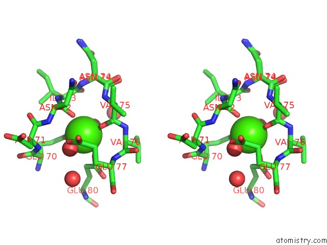
Stereo pair view
Mono view
Stereo pair view
A full contact list of Calcium with other atoms in the Ca binding
site number 1 of Isotropic Trypsin Model For Comparison of Diffuse Scattering within 5.0Å range:
|
Reference:
A.H.Van Benschoten,
L.Liu,
A.Gonzalez,
A.S.Brewster,
N.K.Sauter,
J.S.Fraser,
M.E.Wall.
Measuring and Modeling Diffuse Scattering in Protein X-Ray Crystallography. Proc.Natl.Acad.Sci.Usa V. 113 4069 2016.
ISSN: ESSN 1091-6490
PubMed: 27035972
DOI: 10.1073/PNAS.1524048113
Page generated: Sun Jul 14 19:07:12 2024
ISSN: ESSN 1091-6490
PubMed: 27035972
DOI: 10.1073/PNAS.1524048113
Last articles
Zn in 9J0NZn in 9J0O
Zn in 9J0P
Zn in 9FJX
Zn in 9EKB
Zn in 9C0F
Zn in 9CAH
Zn in 9CH0
Zn in 9CH3
Zn in 9CH1